


ZAP70 Mouse Monoclonal Antibody(C3786)
 Datasheet
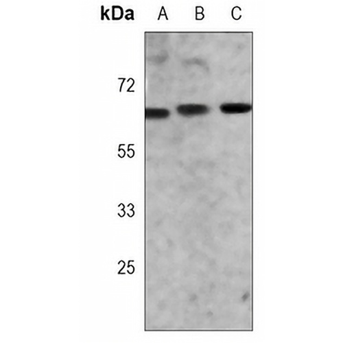
Key features and details
- Target:
- Source/Host:
- Reactivity:
- Clonality:
- Applications:
- Conjugation:
- Storage:
-
Brand:

 Product Details
WB | 1:500 - 1:1000 |
IF/ICC | 1:10 - 1:50 |
FC | 1:10 - 1:50 |
Description | Mouse monoclonal antibody to ZAP70 |
Specificity | Recognizes endogenous levels of ZAP70 protein. |
Antibody Type | Primary antibody |
Imnunogen | Recombinant fusion protein of human ZAP70. The exact sequence is proprietary. |
Purification | This antibody is purified through a protein G column. |
Molecular Weight | Predicted: 69 kD; Observed: 70 kD |
Form/Buffer | Mouse IgG2a kappa. Liquid in PBS, pH 7.3, 30% glycerol, and 0.01% sodium azide. |
Alternative Names | SRK; Tyrosine-protein kinase ZAP-70; 70 kDa zeta-chain associated protein; Syk-related tyrosine kinase |
Gene Symbol | ZAP70 |
Entrez Gene | 7535(Human); 22637(Mouse) |
SwissProt | P43403(Human); P43404(Mouse) |
*Clone Number, Reactivity, Source/Host and Clonality can be found in the product name and Key Features section above.
Western blot analysis of ZAP70 expression in Jurkat (A), MOLT4 (B), mouse thymus (C) whole cell lysates. (Predicted band size: 69 kD; Observed band size: 70 kD)
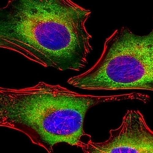
Immunofluorescent analysis of ZAP70 staining in Hela cells. Formalin-fixed cells were permeabilized with 0.1% Triton X-100 in TBS for 5-10 minutes and blocked with 3% BSA-PBS for 30 minutes at room temperature. Cells were probed with the primary antibody in 3% BSA-PBS and incubated overnight at 4 °C in a humidified chamber. Cells were washed with PBST and incubated with a AREX® Fluor 488 -conjugated secondary antibody (green) in PBS at room temperature in the dark. Phalloidin - AREX® Fluor 555 was used to stain Actin filaments (red). DAPI was used to stain the cell nuclei (blue).
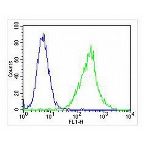
Flow cytometric analysis of Jurkat cells using Anti-ZAP70 Antibody. The cells were fixed with 2% paraformaldehyde (10 min) and then permeabilized with 90% methanol for 10 min. The cells were incubated in 2% bovine serum albumin to block non-specific protein-protein interactions followed by the antibody at 37 °C for 60 min. The secondary antibody Goat Anti-Mouse IgG (H&L) - AREX® Fluor 488 was incubated at 37 °C for 40 min. Isotype control antibody (blue line) was used under the same condition.
FAQs
 New Products


