


WT1 Rabbit Monoclonal Antibody(ARB572)
 Datasheet
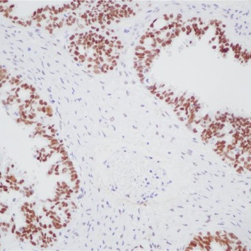
Key features and details
- Target:
- Clone ID:
- Source/Host:
- Reactivity:
- Applications:
- Dilution:
- Clonality:
- Storage:
-
Brand:

CAT.NO. : ARB6864
US$ Please choose
US$ Please choose
Size:
Trail, Bulk size or Custom requests Please contact us
 Product Details
Background
The WT1 gene located at chromosome 11p13 codes for a transcription factor, a DNA - binding nucleoprotein, that plays a role primarily in the development of genitourinary organs. There are at least eight isoforms ranging between 52 and 62 kDa produced by combination of alternative splicing and RNA editing. WT1 is synthesized and reside in the cytoplasm in an inactive form. When activated through phosphorylation it is translocated to the nucleus. WT1 influences cell proliferation by suppressing bcl - 2 and regulating cadherin and p53.In normal epithelia, nuclear WT1 expression is largely restricted to ovary (surface epithelium and inclusion cysts) and fallopian tube, while WT1 is not found in endometrial or cervical epithelium. As regards nonepithelial cells, nuclear WT1 is found in mesothelium and some submesothelial stromal cells, stromal cells of the female genital tract, testicular non - germinal cells, and kidney (podocytes). In tumor tissues, WT1 is detected in tumor cells of Wilms’ Tumor (also known as nephroblastoma) and mesothelioma. Additionally, WT1 expression has been found in ovarian serous carcinomas and some breast carcinomas. WT1 is particularly used for distinguishing malignant mesothelioma and ovarian serous carcinoma from nonserous carcinomas. As for malignant mesothelioma, calretinin and WT1 are superior to cytokeratin 5/6, N - cadherin and thrombomodulin. WT1 is also applicable for the differential diagnostic of small childhood tumors.
Application
To ensure optimal assay performance, AREX recommends conducting reagent titration tailored to each testing system for optimal detection results.
*Results are sample-specific. Please refer to your local assay conditions and test parameters for reference.
Application | Dilution Ratio |
IHC | 1:100 - 1:200 |
Overview
Predicted Molecular Wt | 49kDa |
Purity | ProA affinity purified IgG |
Subcellular location | Nucleus |
Swissprot ID | P19544 |
Immunogen | Synthetic peptide corresponding to WT1 residues within aa1 - 100 of WT1 was used as an immunogen |
Storage Buffer | PBS 59%, Sodium azide 0.01%, Glycerol 40%, BSA 0.05% |
Recommended method | Heat induced epitope retrieval with Tris-EDTA buffer (pH 9.0), primary antibody incubate at RT (18℃-25℃) for 30 minutes |
Alternative Names | Wilms tumor protein 1 |
Gene Symbol | WT1 |
Entrez Gene | 7490 |
*Clone Number, Reactivity, Source/Host and Clonality can be found in the product name and Key Features section above.
Data
Immunohistochemical staining of human ovarian carcinoma tissue using WT1 Rabbit Monoclonal Antibody(ARB572).
Storage
Store at 4°C short term. For long term storage, store at -20°C, avoiding freeze/thaw cycles.
Note
For Research Use Only. Not for diagnostic, therapeutics, prophylactic or in vivo use.
FAQs
Are the pathology antibodies provided by AREX raw antibodies or ready-to-use solutions?

AREX Biosciences specializes in supplying high-quality IHC pathology raw antibodies (Raw Antibodies). We do not manufacture ready-to-use working solutions or IVD diagnostic reagents. We mainly provide concentrated raw materials to pathology reagent manufacturers and diagnostic platform companies to support product development and OEM production.
What development scenarios are pathology raw antibodies mainly used for?
Our raw antibodies are primarily used for the research, performance optimization, validation, and commercial-scale production of pathology IHC detection reagents. They are also suitable for companion diagnostics (CDx) projects and antibody screening.
How can I evaluate whether a raw antibody is suitable for our staining platform?
It is recommended to focus on specificity, sensitivity, background control, and performance on your target automated platforms (such as Roche Ventana, Leica Bond, Agilent Dako, etc.). We can provide internal test data for reference, but we recommend partners perform actual validation on their own platforms.
Can you provide samples for platform validation?
Yes. We can provide validation samples depending on the specific product. Some products may be provided free of charge, while others may involve a small sample fee. Handling fee and shipping fee will be charged separately. Please contact us for detailed confirmation.
How should raw antibodies be paired with detection systems?
AREX raw antibodies can be used with various polymer detection systems and ancillary reagents. We recommend optimization in combination with the ARExVisual® system or your existing detection platform to achieve better signal intensity and background control.
New Products


