


TRAP220 (Phospho-T1457) Rabbit Polyclonal Antibody
 Datasheet
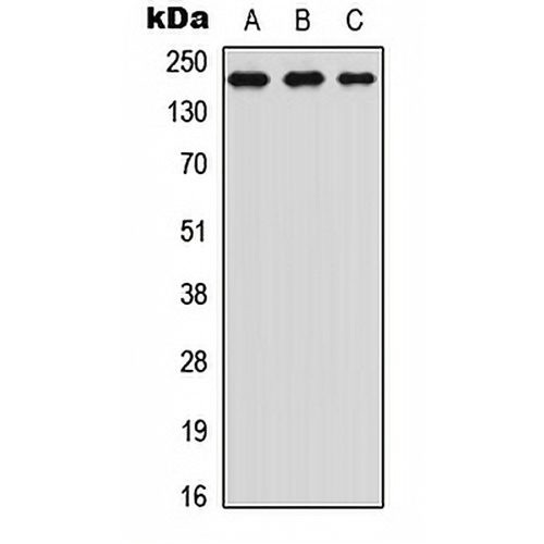
Key features and details
- Target:
- Source/Host:
- Reactivity:
- Clonality:
- Applications:
- Conjugation:
- Storage:
-
Brand:

 Product Details
WB | 1:500 - 1:1000 |
IHC | 1:100 - 1:200 |
IF/ICC | 1:100 - 1:500 |
IP | 1:10 - 1:100 |
ChIP | Use at an assay dependent concentration |
Description | Rabbit polyclonal antibody to TRAP220 (Phospho-T1457) |
Specificity | Recognizes endogenous levels of TRAP220 protein only when phosphorylated at T1457. |
Antibody Type | Primary antibody |
Imnunogen | KLH-conjugated synthetic phosphopeptide corresponding to residues surrounding T1457 of human TRAP220 protein. The exact sequence is proprietary. |
Purification | The antibody was purified by immunogen affinity chromatography. |
Molecular Weight | Predicted: 168 kD; Observed: 260 kD |
Form/Buffer | Liquid in 0.42% Potassium phosphate, 0.87% Sodium chloride, pH 7.3, 30% glycerol, and 0.01% sodium azide. |
Alternative Names | ARC205; CRSP1; CRSP200; DRIP205; DRIP230; PBP; PPARBP; PPARGBP; RB18A; TRAP220; TRIP2; Mediator of RNA polymerase II transcription subunit 1; Activator-recruited cofactor 205 kDa component; ARC205; Mediator complex subunit 1; Peroxisome proliferator-activated receptor-binding protein; PBP; PPAR-binding protein; Thyroid hormone receptor-associated protein complex 220 kDa component; Trap220; Thyroid receptor-interacting protein 2; TR-interacting protein 2; TRIP-2; Vitamin D receptor-interacting protein complex component DRIP205; p53 regulatory protein RB18A |
Gene Symbol | MED1 |
Entrez Gene | 5469(Human); 19014(Mouse) |
SwissProt | Q15648(Human); Q925J9(Mouse) |
*Clone Number, Reactivity, Source/Host and Clonality can be found in the product name and Key Features section above.
Western blot analysis of TRAP220 (Phospho-T1457) expression in Jurkat (A), HeLa (B), NIH3T3 (C) whole cell lysates. (Predicted band size: 168 kD; Observed band size: 260 kD)
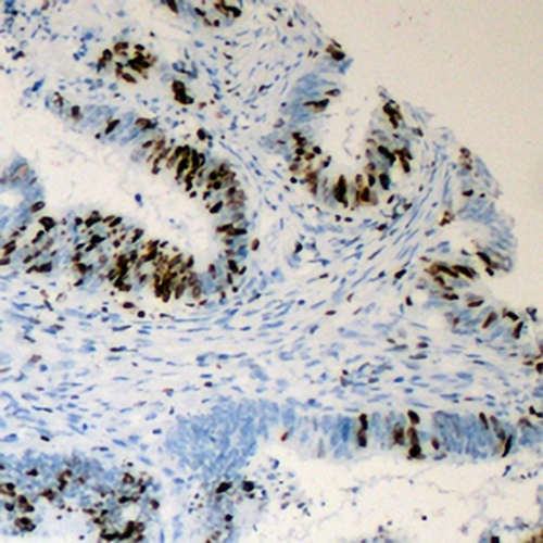
Immunohistochemical analysis of TRAP220 (Phospho-T1457) staining in human colon cancer formalin fixed paraffin embedded tissue section. The section was pre-treated using heat mediated antigen retrieval with sodium citrate buffer (pH 6.0). The section was then incubated with the antibody at room temperature and detected using an HRP conjugated compact polymer system. DAB was used as the chromogen. The section was then counterstained with haematoxylin and mounted with DPX.
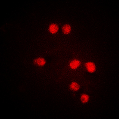
Immunofluorescent analysis of TRAP220 (Phospho-T1457) staining in Jurkat cells. Formalin-fixed cells were permeabilized with 0.1% Triton X-100 in TBS for 5-10 minutes and blocked with 3% BSA-PBS for 30 minutes at room temperature. Cells were probed with the primary antibody in 3% BSA-PBS and incubated overnight at 4 °C in a hidified chamber. Cells were washed with PBST and incubated with a DyLight 594-conjugated secondary antibody (red) in PBS at room temperature in the dark.
FAQs
 New Products


