


Tachykinin Receptor 1 Rabbit Polyclonal Antibody
 Datasheet
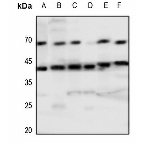
Key features and details
- Target:
- Source/Host:
- Reactivity:
- Clonality:
- Applications:
- Conjugation:
- Storage:
-
Brand:

 Product Details
WB | 1:500 - 1:1000 |
IF/ICC | 1:100 - 1:500 |
Description | Rabbit polyclonal antibody to Tachykinin Receptor 1 |
Specificity | Recognizes endogenous levels of Tachykinin Receptor 1 protein. |
Antibody Type | Primary antibody |
Imnunogen | KLH-conjugated synthetic peptide encompassing a sequence within the center region of human Tachykinin Receptor 1. The exact sequence is proprietary. |
Purification | The antibody was purified by immunogen affinity chromatography. |
Molecular Weight | Predicted: 46 kD; Observed: 46; 53 kD |
Form/Buffer | Liquid in 0.42% Potassium phosphate, 0.87% Sodium chloride, pH 7.3, 30% glycerol, and 0.01% sodium azide. |
Alternative Names | NK1R; TAC1R; Substance-P receptor; SPR; NK-1 receptor; NK-1R; Tachykinin receptor 1 |
Gene Symbol | TACR1 |
Entrez Gene | 6869(Human); 21336(Mouse); 24807(Rat) |
SwissProt | P25103(Human); P30548(Mouse); P14600(Rat) |
*Clone Number, Reactivity, Source/Host and Clonality can be found in the product name and Key Features section above.
Western blot analysis of Tachykinin Receptor 1 expression in HEK293T (A), A2780 (B), mouse spleen (C), mouse kidney (D), rat spleen (E), rat kidney (F) whole cell lysates. (Predicted band size: 46 kD; Observed band size: 46; 53 kD)
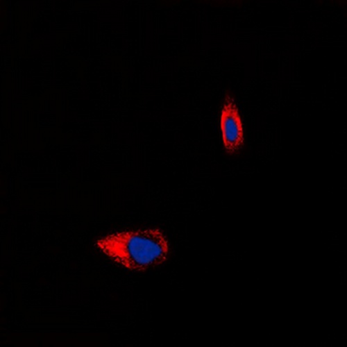
Immunofluorescent analysis of Tachykinin Receptor 1 staining in HeLa cells. Formalin-fixed cells were permeabilized with 0.1% Triton X-100 in TBS for 5-10 minutes and blocked with 3% BSA-PBS for 30 minutes at room temperature. Cells were probed with the primary antibody in 3% BSA-PBS and incubated overnight at 4 °C in a hidified chamber. Cells were washed with PBST and incubated with a DyLight 594-conjugated secondary antibody (red) in PBS at room temperature in the dark. DAPI was used to stain the cell nuclei (blue).
FAQs
 New Products


