


STAT6 Rabbit Monoclonal Antibody(ARB552)
 Datasheet
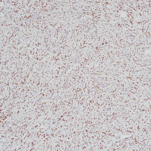
Key features and details
- Target:
- Clone ID:
- Source/Host:
- Reactivity:
- Applications:
- Dilution:
- Clonality:
- Storage:
-
Brand:

CAT.NO. : ARB6844
US$ Please choose
US$ Please choose
Size:
Trail, Bulk size or Custom requests Please contact us
 Product Details
Background
STAT6, a member of the signal transducers and activators of transcription (STAT) family, has been found to form recurrent fusions with NAB2 on chromosome 12q13 in the majority of solitary fibrous tumors. Inactivated STAT6 can be found in the form of a dimer located in the cytoplasm. STAT6 and NAB2 fusion enables cytoplasmic STAT6 to migrate to the nucleus and thus allowing for detection in immunohistochemical assays. NAB2-STAT6 fusion transcripts have been reported in the majority of solitary fibrous tumors but not in meningiomas, hemangioblastomas, schwannomas, and hemangiomas. This makes STAT6 a useful marker in distinguishing solitary fibrous tumors from other morphologically similar tumors.
Application
To ensure optimal assay performance, AREX recommends conducting reagent titration tailored to each testing system for optimal detection results.
*Results are sample-specific. Please refer to your local assay conditions and test parameters for reference.
Application | Dilution Ratio |
IHC | 1:100 - 1:200 |
Overview
Predicted Molecular Wt | 94kDa |
Purity | ProA affinity purified IgG |
Subcellular location | Nucleus/Cytoplasm |
Swissprot ID | P42226 |
Immunogen | Synthetic peptide residues in human STAT6 was used as an immunogen |
Storage Buffer | PBS 59%, Sodium azide 0.01%, Glycerol 40%, BSA 0.05% |
Recommended method | Heat induced epitope retrieval with Tris-EDTA buffer (pH 9.0), primary antibody incubate at RT (18℃-25℃) for 30 minutes |
Alternative Names | Signal transducer and activator of transcription 6; IL-4 Stat |
Gene Symbol | STAT6 |
Entrez Gene | 6778 |
*Clone Number, Reactivity, Source/Host and Clonality can be found in the product name and Key Features section above.
Data
Immunohistochemical staining of human solitary fibrous carcinoma tissue using STAT6 Rabbit Monoclonal Antibody(ARB552).
Storage
Store at 4°C short term. For long term storage, store at -20°C, avoiding freeze/thaw cycles.
Note
For Research Use Only. Not for diagnostic, therapeutics, prophylactic or in vivo use.
FAQs
Are the pathology antibodies provided by AREX raw antibodies or ready-to-use solutions?

AREX Biosciences specializes in supplying high-quality IHC pathology raw antibodies (Raw Antibodies). We do not manufacture ready-to-use working solutions or IVD diagnostic reagents. We mainly provide concentrated raw materials to pathology reagent manufacturers and diagnostic platform companies to support product development and OEM production.
What development scenarios are pathology raw antibodies mainly used for?
Our raw antibodies are primarily used for the research, performance optimization, validation, and commercial-scale production of pathology IHC detection reagents. They are also suitable for companion diagnostics (CDx) projects and antibody screening.
How can I evaluate whether a raw antibody is suitable for our staining platform?
It is recommended to focus on specificity, sensitivity, background control, and performance on your target automated platforms (such as Roche Ventana, Leica Bond, Agilent Dako, etc.). We can provide internal test data for reference, but we recommend partners perform actual validation on their own platforms.
Can you provide samples for platform validation?
Yes. We can provide validation samples depending on the specific product. Some products may be provided free of charge, while others may involve a small sample fee. Handling fee and shipping fee will be charged separately. Please contact us for detailed confirmation.
How should raw antibodies be paired with detection systems?
AREX raw antibodies can be used with various polymer detection systems and ancillary reagents. We recommend optimization in combination with the ARExVisual® system or your existing detection platform to achieve better signal intensity and background control.
New Products


