


Peroxiredoxin 1 Mouse Monoclonal Antibody(C2176)
 Datasheet
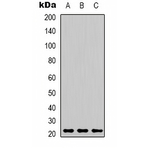
Key features and details
- Target:
- Source/Host:
- Reactivity:
- Clonality:
- Applications:
- Conjugation:
- Storage:
-
Brand:

 Product Details
WB | 1:1000 - 1:3000 |
IF/ICC | 1:100 - 1:200 |
Description | Mouse monoclonal antibody to Peroxiredoxin 1 |
Specificity | Recognizes endogenous levels of Peroxiredoxin 1 protein. |
Antibody Type | Primary antibody |
Imnunogen | Recombinant protein corresponding to human Peroxiredoxin 1. |
Purification | Affinity chromatography |
Molecular Weight | Predicted: 22 kD; Observed: 21 kD |
Form/Buffer | Liquid in 0.42% Potassium phosphate, 0.87% Sodium chloride, pH 7.3, 30% glycerol, and 0.01% sodium azide. |
Alternative Names | PAGA; PAGB; TDPX2; Peroxiredoxin-1; Natural killer cell-enhancing factor A; NKEF-A; Proliferation-associated gene protein; PAG; Thioredoxin peroxidase 2; Thioredoxin-dependent peroxide reductase 2 |
Gene Symbol | PRDX1 |
Entrez Gene | 5052(Human); 18477(Mouse); 117254(Rat) |
SwissProt | Q06830(Human); P35700(Mouse); Q63716(Rat) |
*Clone Number, Reactivity, Source/Host and Clonality can be found in the product name and Key Features section above.
Western blot analysis of Peroxiredoxin 1 expression in MCF7 (A), mouse brain (B), rat kidney (C) whole cell lysates. (Predicted band size: 22 kD; Observed band size: 21 kD)
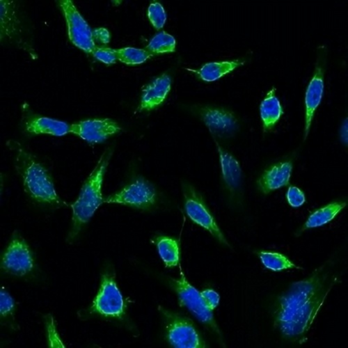
Immunofluorescent analysis of Peroxiredoxin 1 staining in Hela cells. Formalin-fixed cells were permeabilized with 0.1% Triton X-100 in TBS for 5-10 minutes and blocked with 3% BSA-PBS for 30 minutes at room temperature. Cells were probed with the primary antibody in 3% BSA-PBS and incubated overnight at 4 °C in a hidified chamber. Cells were washed with PBST and incubated with a FITC-conjugated secondary antibody (green) in PBS at room temperature in the dark. DAPI was used to stain the cell nuclei (blue).
FAQs
 New Products


