


PDC-E2 Mouse Monoclonal Antibody(C3340)
 Datasheet
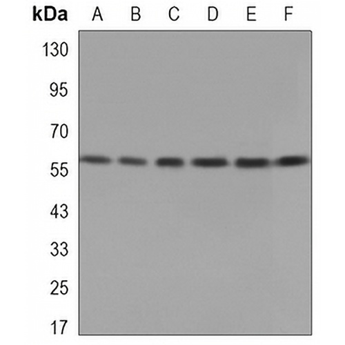
Key features and details
- Target:
- Source/Host:
- Reactivity:
- Clonality:
- Applications:
- Conjugation:
- Storage:
-
Brand:

 Product Details
WB | 1:500 - 1:1000 |
IF/ICC | 1:50 - 1:100 |
IP | 1:10 - 1:50 |
Description | Mouse monoclonal antibody to PDC-E2 |
Specificity | Recognizes endogenous levels of PDC-E2 protein. |
Antibody Type | Primary antibody |
Imnunogen | Purified recombinant human Pyruvate Dehydrogenase E2 protein fragments expressed in E.coli. |
Purification | The antibody was purified by immunogen affinity chromatography. |
Molecular Weight | Predicted: 69 kD; Observed: 69 kD |
Form/Buffer | Liquid in PBS containing 50% glycerol, 0.5% BSA and 0.02% sodium azide, pH 7.3. |
Alternative Names | DLTA; Dihydrolipoyllysine-residue acetyltransferase component of pyruvate dehydrogenase complex, mitochondrial; 70 kDa mitochondrial autoantigen of primary biliary cirrhosis; PBC; Dihydrolipoamide acetyltransferase component of pyruvate dehydrogenase complex; M2 antigen complex 70 kDa subunit; Pyruvate dehydrogenase complex component E2; PDC-E2; PDCE2 |
Gene Symbol | DLAT |
Entrez Gene | 1737(Human); 235339(Mouse) |
SwissProt | P10515(Human); Q8BMF4(Mouse) |
*Clone Number, Reactivity, Source/Host and Clonality can be found in the product name and Key Features section above.
Western blot analysis of PDC-E2 expression in Jurkat (A), A549 (B), U251 (C), F9 (D), Lncap (E), Hela (F) whole cell lysates. (Predicted band size: 69 kD; Observed band size: 69 kD)
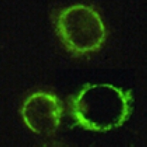
Immunofluorescent analysis of PDC-E2 staining in HeLa cells. Formalin-fixed cells were permeabilized with 0.1% Triton X-100 in TBS for 5-10 minutes and blocked with 3% BSA-PBS for 30 minutes at room temperature. Cells were probed with the primary antibody in 3% BSA-PBS and incubated overnight at 4 °C in a hidified chamber. Cells were washed with PBST and incubated with a AREX® Fluor 488 -conjugated secondary antibody (green) in PBS at room temperature in the dark.
FAQs
 New Products


