p63 Rabbit Monoclonal Antibody(ARB831)

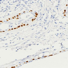
Key features and details

  • Target: p63
  • Clone ID: ARB831
  • Source/Host: Rabbit
  • Reactivity: Human
  • Applications: IHC
  • Dilution: 1:100-1:200
  • Clonality: Monoclonal
  • Storage: at-20°C
  • Brand:
CAT.NO. : ARB6621
US$ Please choose
US$ Please choose
Size:
1mL
Trail, Bulk size or Custom requests Please contact us
Product Details
Background
p63 is a transcription factor related to the tumor suppressor gene p53 and play a critical role in the development of stratified epithelia by maintaining basal cell regenerative capacity and perhaps by playing a role in mediating ectodermal-mesenchymal interactions.In normal tissues, p63 expression is presented in the basal cells of stratified epithelia such as skin, ectocervix, esophagus, urothelium, and bronchi. p63 is also found in basal cells of glandular tissue such as prostate and breast and in lymphoid tissue. In tumor tissues, the p63 gene is amplified and the protein is overexpressed in primary lung and head and neck squamous cell carcinomas. Squamous cell carcinomas of the larynx, esophagus, skin, and cervix also express p63. p63 expression has also been documented in basal cell carcinomas of skin. However, some studies also show that p63 expressed in a subset of adenocarcinomas and large cell carcinomas of lung.In diagnostic pathology practice, p63 has been studied as a marker of basal cells to help in the diagnosis of ductal carcinoma in situ of the breast versus invasive carcinoma in fine-needle aspirates. Similarly, p63 has been tested as a marker to identify basal cells in the diagnostic work-up for prostatic cancer. Recently, some studies have suggested that p63 in combination with other markers may be used to help in differentiating squamous cell carcinomas from other tumor types in cases difficult to diagnose histologically.
Application
To ensure optimal assay performance, AREX recommends conducting reagent titration tailored to each testing system for optimal detection results.

Application

Dilution Ratio

IHC

1:100 - 1:200

*Results are sample-specific. Please refer to your local assay conditions and test parameters for reference.
Overview

Predicted Molecular Wt

77 kDa

Purity

ProA affinity purified IgG

Subcellular location

Nucleus

Swissprot ID

Q9H3D4

Immunogen

Synthetic peptide corresponding to p63 residues within aa580-680 of p63

Storage Buffer

PBS 59%, Sodium azide 0.01%, Glycerol 40%, BSA 0.05%

Recommended method

Heat induced epitope retrieval with Tris-EDTA buffer (pH 9.0), primary antibody incubate at RT (18° C-25° C) for 30 minutes.

Alternative Names

KET; P63; P73H; P73L; TP73L; Tumor protein 63; p63; Chronic ulcerative stomatitis protein; CUSP; Keratinocyte transcription factor KET; Transformation-related protein 63; TP63; Tumor protein p73-like; p73L; p40; p51

Gene Symbol

TP63

Entrez Gene

8626

*AREX continuously optimizes our products. Webpage content may not reflect the latest updates. For inquiries, please contact info@arexbio.com or your local distributor.
*Clone Number, Reactivity, Source/Host and Clonality can be found in the product name and Key Features section above.
Data

Immunohistochemical staining of human prostate tissue sections was performed using p63 Rabbit Monoclonal Antibody (ARB831).

Storage
Store at 4°C short term. For long term storage, store at -20°C, avoiding freeze/thaw cycles.
Note
For Research Use Only. Not for diagnostic, therapeutics, prophylactic or in vivo use.
FAQs
Are the pathology antibodies provided by AREX raw antibodies or ready-to-use solutions?
AREX Biosciences specializes in supplying high-quality IHC pathology raw antibodies (Raw Antibodies). We do not manufacture ready-to-use working solutions or IVD diagnostic reagents. We mainly provide concentrated raw materials to pathology reagent manufacturers and diagnostic platform companies to support product development and OEM production.
What development scenarios are pathology raw antibodies mainly used for?
Our raw antibodies are primarily used for the research, performance optimization, validation, and commercial-scale production of pathology IHC detection reagents. They are also suitable for companion diagnostics (CDx) projects and antibody screening.
How can I evaluate whether a raw antibody is suitable for our staining platform?
It is recommended to focus on specificity, sensitivity, background control, and performance on your target automated platforms (such as Roche Ventana, Leica Bond, Agilent Dako, etc.). We can provide internal test data for reference, but we recommend partners perform actual validation on their own platforms.
Can you provide samples for platform validation?
Yes. We can provide validation samples depending on the specific product. Some products may be provided free of charge, while others may involve a small sample fee. Handling fee and shipping fee will be charged separately. Please contact us for detailed confirmation.
How should raw antibodies be paired with detection systems?
AREX raw antibodies can be used with various polymer detection systems and ancillary reagents. We recommend optimization in combination with the ARExVisual® system or your existing detection platform to achieve better signal intensity and background control.
New Products
Get in touch with AREX
Name:*
Tel/Phone:*
Company:*
Email:*
Inquiry:
Captcha*
Submitting your email information means that you are willing to receive email information from AREX regarding technology, applications, products, and events. By clicking on the 'unsubscribe' button in the email or by contacting info@arexbio.com You can unsubscribe at any time by sending an email. Regarding your data usage information, please refer to our privacy policy.
© AREX 2024. All Rights Reserved. Sitemap
0.172573s