MUC1 Rabbit Monoclonal Antibody(ARB993)

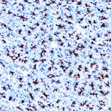
Key features and details

  • Target: MUC1
  • Clone ID: ARB993
  • Source/Host: Rabbit
  • Reactivity: Human
  • Applications: IHC
  • Dilution: 1:100-1:200
  • Clonality: Monoclonal
  • Storage: at-20°C
  • Brand:
CAT.NO. : ARB6785
US$ Please choose
US$ Please choose
Size:
1mL
Trail, Bulk size or Custom requests Please contact us
Product Details
Background
MUC1 (Mucin 1) is a transmembrane glycoprotein that protects epithelial cells from injury caused by external stimuli. In addition to this role, MUC1 is involved in cell-cell adhesion, proliferation, motility, invasion and survival. In epithelial cells, MUC1 expression is regulated by binding of TNFα to TNFR1 and activation of the NFκB pathway.MUC1 is present in a variety of glandular (secretory) epithelia such as breast, eccrine and apocrine glands, and pancreas, whereas little or no MUC1 is expressed in the gastroepithelial epithelium, endocervical epithelium, and prostate glands. The immunoreactivity is usually limited to apical cell membranes, but a staining of the Golgi zone may also be seen. MUC1 can be demonstrated in most types of adenocarcinomas derived from secretory epithelia. In well-differentiated carcinomas, the staining is mostly in apical (apical) cell membranes while in the less differentiated carcinomas, a cytoplasmic staining is seen with loss of staining polarity in the membranes.MUC1 was used to recognize epithelial differentiation when cytokeratin was difficult to visualize. In the classification of hematolymphoid neoplasms, a positive MUC1 reaction may be an aid in the identification of diffuse large B-cell lymphoma, plasma cell neoplasms and nodular lymphocyte predominant Hodgkin's lymphoma.
Application
To ensure optimal assay performance, AREX recommends conducting reagent titration tailored to each testing system for optimal detection results.

Application

Dilution Ratio

IHC

1:100 - 1:200

*Results are sample-specific. Please refer to your local assay conditions and test parameters for reference.
Overview

Predicted Molecular Wt

122kDa

Purity

ProA affinity purified IgG

Subcellular location

Membrane/Cytoplasm

Swissprot ID

P15941

Immunogen

Synthetic peptide corresponding to MUC1 residues within aa1155-1255 of MUC1 was used as an immunogen

Storage Buffer

PBS 59%, Sodium azide 0.01%, Glycerol 40%, BSA 0.05%

Recommended method

Heat induced epitope retrieval with Tris-EDTA buffer (pH 9.0), primary antibody incubate at RT (18℃-25℃) for 30 minutes

Alternative Names

PUM; Mucin-1; MUC-1; Breast carcinoma-associated antigen DF3; Cancer antigen 15-3; CA 15-3; Carcinoma-associated mucin; Episialin; H23AG; Krebs von den Lungen-6; KL-6; PEMT; Peanut-reactive urinary mucin; PUM; Polymorphic epithelial mucin; PEM; Tumor-associated epithelial membrane antigen; EMA; Tumor-associated mucin; CD227

Gene Symbol

MUC1

Entrez Gene

4582

*AREX continuously optimizes our products. Webpage content may not reflect the latest updates. For inquiries, please contact info@arexbio.com or your local distributor.
*Clone Number, Reactivity, Source/Host and Clonality can be found in the product name and Key Features section above.
Data

Immunohistochemical staining of human pancreas tissue using MUC1 Rabbit Monoclonal Antibody(ARB993).

Storage
Store at 4°C short term. For long term storage, store at -20°C, avoiding freeze/thaw cycles.
Note
For Research Use Only. Not for diagnostic, therapeutics, prophylactic or in vivo use.
FAQs
Are the pathology antibodies provided by AREX raw antibodies or ready-to-use solutions?
AREX Biosciences specializes in supplying high-quality IHC pathology raw antibodies (Raw Antibodies). We do not manufacture ready-to-use working solutions or IVD diagnostic reagents. We mainly provide concentrated raw materials to pathology reagent manufacturers and diagnostic platform companies to support product development and OEM production.
What development scenarios are pathology raw antibodies mainly used for?
Our raw antibodies are primarily used for the research, performance optimization, validation, and commercial-scale production of pathology IHC detection reagents. They are also suitable for companion diagnostics (CDx) projects and antibody screening.
How can I evaluate whether a raw antibody is suitable for our staining platform?
It is recommended to focus on specificity, sensitivity, background control, and performance on your target automated platforms (such as Roche Ventana, Leica Bond, Agilent Dako, etc.). We can provide internal test data for reference, but we recommend partners perform actual validation on their own platforms.
Can you provide samples for platform validation?
Yes. We can provide validation samples depending on the specific product. Some products may be provided free of charge, while others may involve a small sample fee. Handling fee and shipping fee will be charged separately. Please contact us for detailed confirmation.
How should raw antibodies be paired with detection systems?
AREX raw antibodies can be used with various polymer detection systems and ancillary reagents. We recommend optimization in combination with the ARExVisual® system or your existing detection platform to achieve better signal intensity and background control.
New Products
Get in touch with AREX
Name:*
Tel/Phone:*
Company:*
Email:*
Inquiry:
Captcha*
Submitting your email information means that you are willing to receive email information from AREX regarding technology, applications, products, and events. By clicking on the 'unsubscribe' button in the email or by contacting info@arexbio.com You can unsubscribe at any time by sending an email. Regarding your data usage information, please refer to our privacy policy.
© AREX 2024. All Rights Reserved. Sitemap
0.206244s