


MNDA Rabbit Polyclonal Antibody
 Datasheet
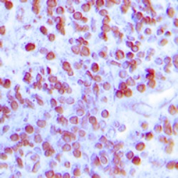
Key features and details
- Target:
- Host:
- Reactivity:
- Clonality:
- Application:
- Storage:
-
Brand:

CAT.NO. : ARA6780
US$ Please choose
US$ Please choose
Size:
Trail, Bulk size or Custom requests Please contact us
 Product Details
Background
The myeloid cell nuclear differentiation antigen (MNDA) is detected only in nuclei of cells of the granulocyte-monocyte lineage. A 200-amino acid region of human MNDA is strikingly similar to a region in the proteins encoded by a family of interferon-inducible mouse genes, designated Ifi-201, Ifi-202, and Ifi-203, that are not regulated in a cell- or tissue-specific fashion. The 1.8-kb MNDA mRNA, which contains an interferon-stimulated response element in the 5-prime untranslated region, was significantly upregulated in human monocytes exposed to interferon alpha. MNDA is located within 2,200 kb of FCER1A, APCS, CRP, and SPTA1. In its pattern of expression and/or regulation, MNDA resembles IFI16, suggesting that these genes participate in blood cell-specific responses to interferons. [provided by RefSeq, Jul 2008]
Application
To ensure optimal assay performance, AREX recommends conducting reagent titration tailored to each testing system for optimal detection results.
*Results are sample-specific. Please refer to your local assay conditions and test parameters for reference.
Application | Dilution Ratio |
IHC | 1:50-1:100 |
Overview
Immunogen | KLH-conjugated synthetic peptide encompassing a sequence within the C-term region of human MNDA. The exact sequence is proprietary. |
Purification | The antibody was purified by immunogen affinity chromatography. |
Form/Buffer | Liquid in 0.42% Potassium phosphate, 0.87% Sodium chloride, pH 7.3, 30% glycerol, and 0.01% sodium azide. |
Gene Name | MNDA |
Related Names | Myeloid cell nuclear differentiation antigen |
Gene ID (Human) | 4332 |
Protein ID (Human) | P41218 |
*Clone Number, Reactivity, Source/Host and Clonality can be found in the product name and Key Features section above.
Data
Immunohistochemical staining of human lymph node tissue using MNDA Rabbit Polyclonal Antibody
Storage
Shipped at 4℃. Store at -20℃ for one year. Avoid repeated freeze/thaw cycles.
Note
For Research Use Only. Not for diagnostic, therapeutics, prophylactic or in vivo use.
FAQs
Are the pathology antibodies provided by AREX raw antibodies or ready-to-use solutions?

AREX Biosciences specializes in supplying high-quality IHC pathology raw antibodies (Raw Antibodies). We do not manufacture ready-to-use working solutions or IVD diagnostic reagents. We mainly provide concentrated raw materials to pathology reagent manufacturers and diagnostic platform companies to support product development and OEM production.
What development scenarios are pathology raw antibodies mainly used for?
Our raw antibodies are primarily used for the research, performance optimization, validation, and commercial-scale production of pathology IHC detection reagents. They are also suitable for companion diagnostics (CDx) projects and antibody screening.
How can I evaluate whether a raw antibody is suitable for our staining platform?
It is recommended to focus on specificity, sensitivity, background control, and performance on your target automated platforms (such as Roche Ventana, Leica Bond, Agilent Dako, etc.). We can provide internal test data for reference, but we recommend partners perform actual validation on their own platforms.
Can you provide samples for platform validation?
Yes. We can provide validation samples depending on the specific product. Some products may be provided free of charge, while others may involve a small sample fee. Handling fee and shipping fee will be charged separately. Please contact us for detailed confirmation.
How should raw antibodies be paired with detection systems?
AREX raw antibodies can be used with various polymer detection systems and ancillary reagents. We recommend optimization in combination with the ARExVisual® system or your existing detection platform to achieve better signal intensity and background control.
New Products


