


Histone Deacetylase 7 (Phospho-S155) Rabbit Polyclonal Antibody
 Datasheet
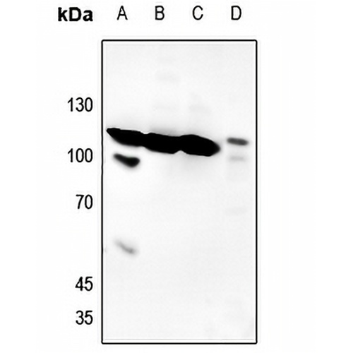
Key features and details
- Target:
- Source/Host:
- Reactivity:
- Clonality:
- Applications:
- Conjugation:
- Storage:
-
Brand:

 Product Details
WB | 1:500 - 1:1000 |
IF/ICC | 1:100 - 1:500 |
Description | Rabbit polyclonal antibody to Histone Deacetylase 7 (Phospho-S155) |
Specificity | Recognizes endogenous levels of Histone Deacetylase 7 protein only when phosphorylated at S155. |
Antibody Type | Primary antibody |
Imnunogen | KLH-conjugated synthetic phosphopeptide corresponding to residues surrounding S155 of human Histone Deacetylase 7 protein. The exact sequence is proprietary. |
Purification | The antibody was purified by immunogen affinity chromatography. |
Molecular Weight | Predicted: 102 kD; Observed: 105 kD |
Form/Buffer | Liquid in 0.42% Potassium phosphate, 0.87% Sodium chloride, pH 7.3, 30% glycerol, and 0.01% sodium azide. |
Alternative Names | HDAC7A; Histone deacetylase 7; HD7; Histone deacetylase 7A; HD7a |
Gene Symbol | HDAC7 |
Entrez Gene | 51564(Human); 56233(Mouse) |
SwissProt | Q8WUI4(Human); Q8C2B3(Mouse); Q99P96(Rat) |
*Clone Number, Reactivity, Source/Host and Clonality can be found in the product name and Key Features section above.
Western blot analysis of Histone Deacetylase 7 (Phospho-S155) expression in HEK293T (A), Hela (B), U2OS (C), rat lung (D) whole cell lysates. (Predicted band size: 102 kD; Observed band size: 105 kD)

Immunofluorescent analysis of Histone Deacetylase 7 (Phospho-S155) staining in HepG2 cells. Formalin-fixed cells were permeabilized with 0.1% Triton X-100 in TBS for 5-10 minutes and blocked with 3% BSA-PBS for 30 minutes at room temperature. Cells were probed with the primary antibody in 3% BSA-PBS and incubated overnight at 4 °C in a hidified chamber. Cells were washed with PBST and incubated with a DyLight 594-conjugated secondary antibody (red) in PBS at room temperature in the dark.
FAQs
 New Products


