G-CSF Rabbit Monoclonal Antibody(C4068)


Key features and details
- Reactivity:
- Application:
- Host:
- Clonality:
- Target:
-
Brand:

CAT.NO. : AMA03888
US$ Please choose
US$ Please choose
 Product Details
Background
G-CSF (Granulocyte colony stimulating factor) is a naturally occurring cytokine that stimulates the production and antibacterial function of neutrophils and monocytes. Human G-CSF is an 18.8 kDa protein containing 175 amino acid residues, and a soluble isoform of the G-CSF receptor has been described. The pleotropic cytokine is produced by activated monocytes, macrophages, endothelial cells, fibroblasts, astrocytes, osteoblasts and bone marrow cells. G-CSF has been shown to have specific effects on the proliferation, differentiation and activation of hematopoietic cells. G-CSF is also expressed by various transformed cells such as carcinoma cells and myeloblastic leukemia cells. G-CSF is encoded by two distinct DNA sequences, resulting in a full size, high activity and a shorter, low activity isoform of G-CSF. G-CSF is highly conserved among species and has been shown to exert its biological functions through interaction with its receptor expressed on the surface of hematopoietic progenitors, neutrophilic granulocytes and certain carcinoma cell lines. Clinical use of G-CSF has been approved for several therapeutic applications, treatment of neonatal infections, therapy of acute myocardial infarction, granulocyte transfusion in patients with neutropenia, in severe infections and sepsis, therapy in chronic autoimmune neutropenia, treatment of acute myeloid leukemias, Sweet's syndrome and AIDS. Further, G-CSF has been shown to be a marker protein for different carcinomas such as bladder cancer and dysfunction of the protein has been linked to Kostmann Syndrome.
Application
|
Application |
Dilution Ratio |
|
WB |
1:500-1:2000 |
|
IF/ICC |
1:50-1:200 |
Overview
|
Product Description |
Recombinant rabbit monoclonal antibody to G-CSF |
|
Immunogen |
KLH-conjugated synthetic peptide encompassing a sequence within human G-CSF protein. The exact sequence is proprietary. |
|
Purification Method |
The antibody was purified by immunogen affinity chromatography. |
|
Clonality |
Monoclonal |
|
Form |
Liquid in PBS, pH 7.3, 50% glycerol, 0.05% BSA, and 0.05% Proclin300. |
|
Gene Symbol |
CSF3 |
|
Alternative Names |
C17orf33; GCSF; Granulocyte colony-stimulating factor; G-CSF; Pluripoietin; Filgrastim; Lenograstim |
|
Gene ID (Human) |
1440 |
|
Gene ID (Mouse) |
12985 |
|
Protein ID (Human) |
P09919 |
|
Protein ID (Mouse) |
P09920 |
Data

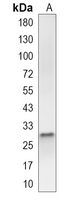
Western blot analysis of G-CSF expression in MCF7 (A) whole cell lysates. (Predicted band size: 22 kD; Observed band size: 30 kD)
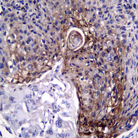
Immunohistochemical analysis of G-CSF staining in human esophagus cancer formalin fixed paraffin embedded tissue section. The section was pre-treated using heat mediated antigen retrieval with sodium citrate buffer (pH 6.0). The section was then incubated with the antibody at room temperature and detected using an HRP conjugated compact polymer system. DAB was used as the chromogen. The section was then counterstained with haematoxylin and mounted with DPX.
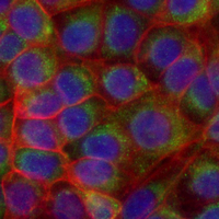
Immunofluorescent analysis of G-CSF staining in MCF7 cells. Formalin-fixed cells were permeabilized with 0.1% Triton X-100 in TBS for 5-10 minutes and blocked with 3% BSA-PBS for 30 minutes at room temperature. Cells were probed with the primary antibody in 3% BSA-PBS and incubated overnight at 4 °C in a hidified chamber. Cells were washed with PBST and incubated with an AREX Flour 488-conjugated secondary antibody (green) in PBS at room temperature in the dark. Phalloidin - AREX Flour 594 was used to stain Actin filaments (red). DAPI was used to stain the cell nuclei (blue).
Storage
Store at 4°C short term. For long term storage, store at -20°C, avoiding freeze/thaw cycles.
Research Use Only
For Research Use Only. Not for use in diagnostic procedures.
New Products