


ERK2 Rabbit Monoclonal Antibody(ARA773)
 Datasheet
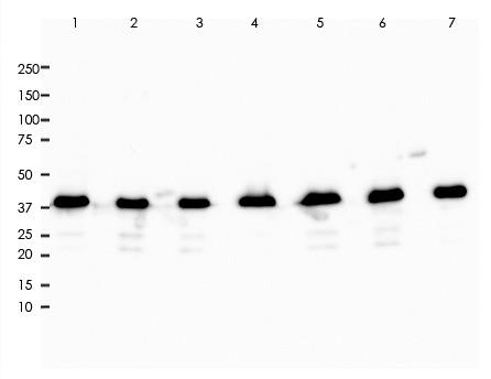
Key features and details
- Target:
- Source/Host:
- Reactivity:
- Clonality:
- Applications:
- Conjugation:
- Storage:
-
Brand:

 Product Details
WB | 1:1000-1:2000 |
IHC | 1:800-1:1600 |
FC | 1:200-1:1000 |
IP | 1:20 |
Description | Rabbit Monoclonal Antibody to ERK2 |
Antibody Type | Primary antibody |
Predicted MW | 41kDa |
Immunogen | A synthetic peptide corresponding to the C-term of ERK2 was used as an immunogen. |
Purification | ProA affinity purified IgG |
Form/Buffer | PBS 59%, Sodium azide 0.01%, Glycerol 40%, BSA 0.48%. |
Alternative Names | MAPK1; ERK2; PRKM1; PRKM2; Mitogen-activated protein kinase 1; MAP kinase 1; MAPK 1; ERT1; Extracellular signal-regulated kinase 2; ERK-2; MAP kinase isoform p42; p42-MAPK; Mitogen-activated protein kinase 2; MAP kinase 2; MAPK 2; MAPK3; ERK1; PRKM3; Mitogen-activated protein kinase 3; MAP kinase 3; MAPK 3; ERT2; Extracellular signal-regulated kinase 1; ERK-1; Insulin-stimulated MAP2 kinase; MAP kinase isoform p44; p44-MAPK; Microtubule-associated protein 2 kinase; p44-ERK1 |
Gene Symbol | MAPK1; MAPK3 |
Entrez Gene | 5594; 5595(Human) |
Swissprot | P28482 |
*Clone Number, Reactivity, Source/Host and Clonality can be found in the product name and Key Features section above.
All lanes: Anti-ERK2 antibody at 1:1,000 dilution
Predicted MW: 41 kDa
Observed MW: 42 kDa
Lane 1: JurKat
Lane 2: Hela
Lane 3: 293
Lane 4: A431
Lane 5: Raw264.7
Lane 6: 3T3
Lane 7: PC-12
Lysate at 10 µg per lane
2nd Ab:
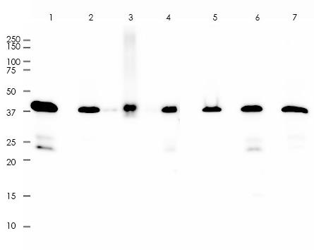
All lanes: Anti-ERK2 antibody at 1:1,000 dilution
Predicted MW: 41 kDa
Observed MW: 42 kDa
Lane 1: Mu Brain
Lane 2: Mu Heart
Lane 3: Mu Kidney
Lane 4: Mu Liver
Lane 5: Rat Heart
Lane 6: Rat Kidney
Lane 7: Rat Liver
Lysate at 10 µg per lane
2nd Ab:
GAR HRP(H+L) 1:10,000
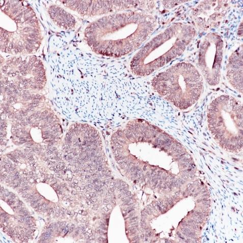
Immunohistochemistry (Formalin/PFA-fixed paraffin-embedded sections) analysis of endometrium cancer tissue labelling ERK2 with ARA773 at 1:1,600. Heat mediated antigen retrieval was performed using Tris/EDTA buffer pH 9.0.
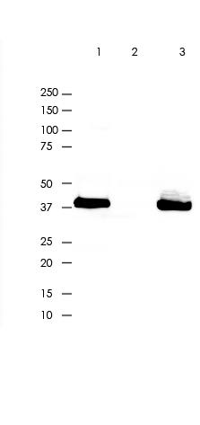
Anti-ERK2 was immunoprecipitated from 0.4mg of A431 lysate with ARA773 at 1:20 dilution.
2nd Ab:
GAR HRP for IP 1:10,000
Lane 1: ARA773 IP in A431 whole cell lysate
Lane 2: PBS instead of ARA773 in A431 whole cell lysate Lane3: A431 whole cell lysate, 10 µg(input)
Exposure: 120s
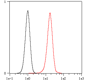
Overlay histogram showing Hela cells stained with ARA773 (Red). The cells were fixed with 4% paraformaldehyde (10 min) and then permeabilized with 0.1% TritonX-100 for 15 min. The cells were then incubated in the antibody (ARA773 , 1:200 dilution) in 1x PBS/1% BSA for 30 min at room temperature . The secondary antibody used was a Goat Anti-Rabbit Alexa Fluor<sup>®</sup> 488 (IgG H+L) at 1:2,000 dilution for 20 min at room temperature . Unlabelled sample (Black) was used as a control.
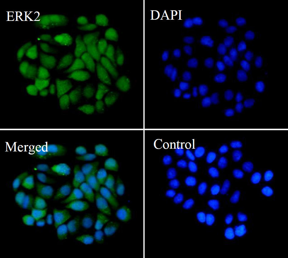
ARA773 staining ERK2 in Hela cells by ICC/IF (Immunocytochemistry/immunofluorescence). Cells were fixed with paraformaldehyde, permeabilized with 0.1% Triton X-100 and blocked with 10% goat serum for half an hour at room temperature. Samples were incubated with primary antibody (1:50) at 4°C. An Alexa Fluor<sup>®</sup> 488-conjugated Goat Anti-Rabbit IgG polyclonal was used as the secondary antibody (1:500). DAPI (blue) was used as the nuclear counter stain.
Control: PBS and secondary antibody, An Alexa Fluor<sup>®</sup> 488-conjugated Goat Anti-Rabbit IgG(1:500).
FAQs
 New Products


