


DDB1 Rabbit Monoclonal Antibody(C1429)
 Datasheet
Key features and details
- Target:
- Source/Host:
- Reactivity:
- Clonality:
- Applications:
- Conjugation:
- Storage:
-
Brand:

 Product Details
WB | 1:500 - 1:1000 |
IHC | 1:50 - 1:200 |
Description | Recombinant rabbit monoclonal antibody to DDB1 |
Specificity | Recognizes endogenous levels of DDB1 protein |
Antibody Type | Primary antibody, Recombinant |
Imnunogen | KLH-conjugated synthetic peptide encompassing a sequence within human DDB1 protein. The exact sequence is proprietary. |
Purification | The antibody was purified by immunogen affinity chromatography. |
Molecular Weight | Predicted: 126 kD; Observed: 127 kD |
Form/Buffer | Liquid in PBS, pH 7.4, containing 50% glycerol, 0.2% BSA and 0.01% sodium azide. |
Alternative Names | XAP1; DNA damage-binding protein 1; DDB p127 subunit; DNA damage-binding protein a; DDBa; Damage-specific DNA-binding protein 1; HBV X-associated protein 1; XAP-1; UV-damaged DNA-binding factor; UV-damaged DNA-binding protein 1; UV-DDB 1; XPE-binding factor; XPE-BF; Xeroderma pigmentosum group E-complementing protein; XPCe |
Gene Symbol | DDB1 |
Entrez Gene | 1642(Human); 13194(Mouse) |
SwissProt | Q16531(Human); Q3U1J4(Mouse); Q9ESW0(Rat) |
*Clone Number, Reactivity, Source/Host and Clonality can be found in the product name and Key Features section above.
Western blot analysis of DDB1 expression in K562 (A), MCF7 (B), mouse kidney (C), mouse muscle (D), rat kidney (E), rat muscle (F) whole cell lysates. (Predicted band size: 126 kD; Observed band size: 127 kD)
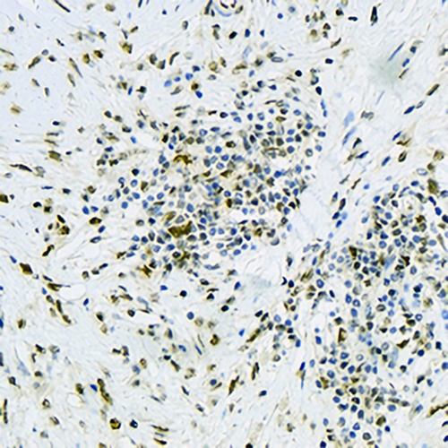
Immunohistochemical analysis of DDB1 staining in human kidney formalin fixed paraffin embedded tissue section. The section was pre-treated using heat mediated antigen retrieval with sodium citrate buffer (pH 6.0). The section was then incubated with the antibody at room temperature and detected using an HRP conjugated compact polymer system. DAB was used as the chromogen. The section was then counterstained with haematoxylin and mounted with DPX.
FAQs
 New Products


