


CD43 Mouse Monoclonal Antibody(ARM949)
 Datasheet
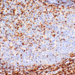
Key features and details
- Target:
- Clone ID:
- Source/Host:
- Reactivity:
- Applications:
- Dilution:
- Clonality:
- Storage:
-
Brand:

CAT.NO. : ARM6739
US$ Please choose
US$ Please choose
Size:
Trail, Bulk size or Custom requests Please contact us
 Product Details
Background
Cluster of Differentiation 43 (CD43), also known as Sialophorin, is a transmembrane protein that plays a role in T-cell activation. CD43 is normally expressed abundantly on the surface of differentiated hematopoietic stem cells, including monocytes, granulocytes, T-lymphocytes, and some B-lymphocytes. Due to the efficacy of CD43 immunohistochemical staining in granulocytes, it is an effective marker for myeloid tumours, while other antibodies demonstrate weak staining under these conditions. Given the low reactivity of Anti-CD43 with B-cells, positive staining of CD43 is implicated in a number of lymphoid and myeloid tumours, with over 90% positive staining in T-cell lymphomas. When CD43 is used in combination with CD45 and L26, immunotyping of various lymphomas can be obtained; this is particularly true when co staining a lymphoid infiltrate with CD20 and CD3.
Application
To ensure optimal assay performance, AREX recommends conducting reagent titration tailored to each testing system for optimal detection results.
*Results are sample-specific. Please refer to your local assay conditions and test parameters for reference.
Application | IHC |
Dilution Ratio | 1:50 - 1:200 |
Overview
Antibody Type | Primary antibodies |
Isotype | IgG1 |
Positive Control | Tonsil tissues, Lymph Node |
Localization | Membranous |
Form/Buffer | Tris Buffer, pH 7.3 - 7.7, with 1% BSA and <0.1% Sodium Azide |
Purification | Purified |
Conjugation | Unconjugated |
Alternative Names | CD43; Leukosialin; Galactoglycoprotein; GALGP; Leukocyte sialoglycoprotein; Sialophorin; CD43 |
Gene Symbol | SPN |
Entrez Gene | 6693 |
Uniprot | P16150 |
*Clone Number, Reactivity, Source/Host and Clonality can be found in the product name and Key Features section above.
Data
Immunohistochemical staining of human tonsil tissue using CD43 Mouse Monoclonal Antibody (ARM949).
Research Use Only
For Research Use Only. Not for diagnostic, therapeutics, prophylactic or in vivo use.
Storage
Store at 4°C short term. For long term storage, store at -20°C, avoiding freeze/thaw cycles.
FAQs
Are the pathology antibodies provided by AREX raw antibodies or ready-to-use solutions?

AREX Biosciences specializes in supplying high-quality IHC pathology raw antibodies (Raw Antibodies). We do not manufacture ready-to-use working solutions or IVD diagnostic reagents. We mainly provide concentrated raw materials to pathology reagent manufacturers and diagnostic platform companies to support product development and OEM production.
What development scenarios are pathology raw antibodies mainly used for?
Our raw antibodies are primarily used for the research, performance optimization, validation, and commercial-scale production of pathology IHC detection reagents. They are also suitable for companion diagnostics (CDx) projects and antibody screening.
How can I evaluate whether a raw antibody is suitable for our staining platform?
It is recommended to focus on specificity, sensitivity, background control, and performance on your target automated platforms (such as Roche Ventana, Leica Bond, Agilent Dako, etc.). We can provide internal test data for reference, but we recommend partners perform actual validation on their own platforms.
Can you provide samples for platform validation?
Yes. We can provide validation samples depending on the specific product. Some products may be provided free of charge, while others may involve a small sample fee. Handling fee and shipping fee will be charged separately. Please contact us for detailed confirmation.
How should raw antibodies be paired with detection systems?
AREX raw antibodies can be used with various polymer detection systems and ancillary reagents. We recommend optimization in combination with the ARExVisual® system or your existing detection platform to achieve better signal intensity and background control.
New Products


