


CD326 Rabbit Monoclonal Antibody(C3144)
 Datasheet
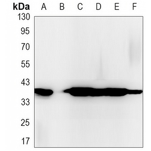
Key features and details
- Target:
- Source/Host:
- Reactivity:
- Clonality:
- Applications:
- Conjugation:
- Storage:
-
Brand:

 Product Details
WB | 1:500 - 1:1000 |
IHC | 1:50 - 1:100 |
IF/ICC | 1:50 - 1:100 |
IP | 1:10 - 1:50 |
Description | Rabbit monoclonal antibody to CD326 |
Specificity | Recognizes endogenous levels of CD326 protein. |
Antibody Type | Primary antibody |
Imnunogen | Recombinant protein of human EpCAM |
Purification | The antibody was purified by immunogen affinity chromatography. |
Molecular Weight | Predicted: 35 kD; Observed: 35; 39 kD |
Form/Buffer | Liquid in 50mM Tris-Glycine (pH 7.4), 0.15M NaCl, 50% Glycerol, 0.01% Sodium azide and 0.05% BSA. |
Alternative Names | GA733-2; M1S2; M4S1; MIC18; TACSTD1; TROP1; Epithelial cell adhesion molecule; Ep-CAM; Adenocarcinoma-associated antigen; Cell surface glycoprotein Trop-1; Epithelial cell surface antigen; Epithelial glycoprotein; EGP; Epithelial glycoprotein 314; EGP314; hEGP314; KS 1/4 antigen; KSA; Major gastrointestinal tumor-associated protein GA733-2; Tumor-associated calcium signal transducer 1; CD326 |
Gene Symbol | EPCAM |
Entrez Gene | 4072(Human); 17075(Mouse); 171577(Rat) |
SwissProt | P16422(Human); Q99JW5(Mouse); O55159(Rat) |
*Clone Number, Reactivity, Source/Host and Clonality can be found in the product name and Key Features section above.
Western blot analysis of CD326 expression in MCF7 (A), HCT116 (B), mouse colon (C) whole cell lysates. (Predicted band size: 35 kD; Observed band size: 35; 39 kD)
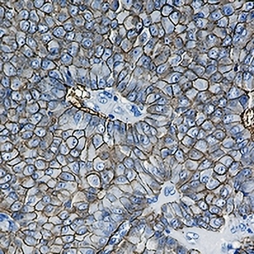
Immunohistochemical analysis of CD326 staining in human breast carcinoma formalin fixed paraffin embedded tissue section. The section was pre-treated using heat mediated antigen retrieval with sodium citrate buffer (pH 6.0). The section was then incubated with the antibody at room temperature and detected using an HRP conjugated compact polymer system. DAB was used as the chromogen. The section was then counterstained with haematoxylin and mounted with DPX.
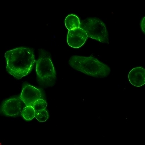
Immunofluorescent analysis of CD326 staining in MCF7 cells. Formalin-fixed cells were permeabilized with 0.1% Triton X-100 in TBS for 5-10 minutes and blocked with 3% BSA-PBS for 30 minutes at room temperature. Cells were probed with the primary antibody in 3% BSA-PBS and incubated overnight at 4 °C in a hidified chamber. Cells were washed with PBST and incubated with a AREX® Fluor 488 -conjugated secondary antibody (green) in PBS at room temperature in the dark.
FAQs
 New Products


