


CD322 Mouse Monoclonal Antibody(C2785)
 Datasheet
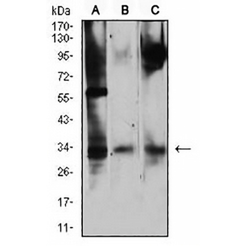
Key features and details
- Target:
- Source/Host:
- Reactivity:
- Clonality:
- Applications:
- Conjugation:
- Storage:
-
Brand:

 Product Details
WB | 1:500 - 1:1000 |
IF/ICC | 1:50 - 1:100 |
FC | 1:100 - 1:200 |
Description | Mouse monoclonal to CD322 |
Specificity | Recognizes endogenous levels of CD322 protein |
Antibody Type | Primary antibody |
Imnunogen | Recombinant fusion protein of human CD322 expressed in E. Coli |
Purification | This antibody is purified through a protein G column. |
Molecular Weight | Predicted: 33 kD; Observed: 33 kD kD |
Form/Buffer | Mouse IgG1. Liquid in PBS, pH 7.3, 30% glycerol, and 0.01% sodium azide. |
Alternative Names | C21orf43; VEJAM; Junctional adhesion molecule B; JAM-B; Junctional adhesion molecule 2; JAM-2; Vascular endothelial junction-associated molecule; VE-JAM; CD322 |
Gene Symbol | JAM2 |
Entrez Gene | 58494(Human) |
SwissProt | P57087(Human) |
*Clone Number, Reactivity, Source/Host and Clonality can be found in the product name and Key Features section above.
Western blot analysis of CD322 expression in NIH/3T3 (A), Ramos (B), HepG2 (C) whole cell lysates. (Predicted band size: 33 kD; Observed band size: 33 kD kD)
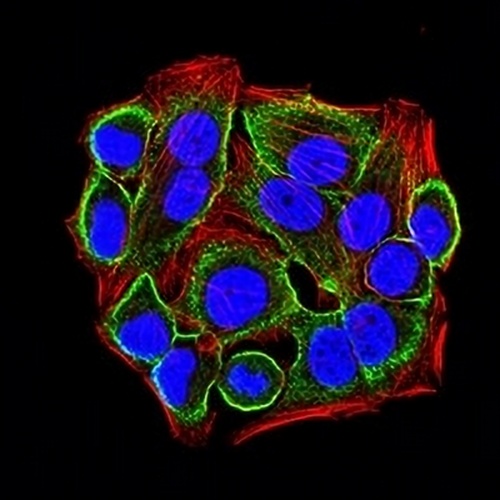
Immunofluorescent analysis of CD322 staining in Hela cells. Formalin-fixed cells were permeabilized with 0.1% Triton X-100 in TBS for 5-10 minutes and blocked with 3% BSA-PBS for 30 minutes at room temperature. Cells were probed with the primary antibody in 3% BSA-PBS and incubated overnight at 4 °C in a hidified chamber. Cells were washed with PBST and incubated with an AREX® Fluor 488 -conjugated secondary antibody (green) in PBS at room temperature in the dark. Phalloidin - AREX® Fluor 594 was used to stain Actin filaments (red). DAPI was used to stain the cell nuclei (blue).
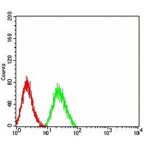
Flow cytometric analysis of HL60 cells using Anti-CD322 Antibody (green) and negative control (red).
FAQs
 New Products


