


Caspase 8 Mouse Monoclonal Antibody(C3904)
 Datasheet
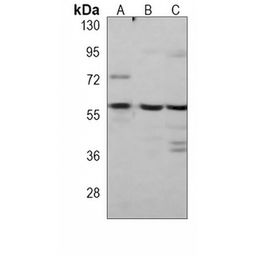
Key features and details
- Target:
- Source/Host:
- Reactivity:
- Clonality:
- Applications:
- Conjugation:
- Storage:
-
Brand:

 Product Details
WB | 1:500 - 1:1000 |
FC | 1:10 - 1:50 |
Description | Mouse monoclonal antibody to Caspase 8 |
Specificity | Recognizes endogenous levels of Caspase 8 protein. |
Antibody Type | Primary antibody |
Imnunogen | KLH-conjugated synthetic peptide encompassing a sequence within the C-term region of human Caspase 8. The exact sequence is proprietary. |
Purification | This antibody is purified through a protein G column. |
Molecular Weight | Predicted: 55 kD; Observed: 60 kD |
Form/Buffer | Mouse IgG1. Liquid in PBS, pH 7.3, 30% glycerol, and 0.01% sodium azide. |
Alternative Names | MCH5; Caspase-8; CASP-8; Apoptotic cysteine protease; Apoptotic protease Mch-5; CAP4; FADD-homologous ICE/ced-3-like protease; FADD-like ICE; FLICE; ICE-like apoptotic protease 5; MORT1-associated ced-3 homolog; MACH |
Gene Symbol | CASP8 |
Entrez Gene | 841(Human) |
SwissProt | Q14790(Human) |
*Clone Number, Reactivity, Source/Host and Clonality can be found in the product name and Key Features section above.
Western blot analysis of Caspase 8 expression in HepG2 (A), Jurkat (B), HL60 (C) whole cell lysates. (Predicted band size: 55 kD; Observed band size: 60 kD)
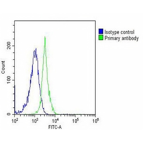
Flow cytometric analysis of Jurkat cells using Anti-Caspase 8 Antibody. The cells were fixed with 2% paraformaldehyde (10 min) and then permeabilized with 90% methanol for 10 min. The cells were incubated in 2% bovine serum albumin to block non-specific protein-protein interactions followed by the antibody at 37 °C for 60 min. The secondary antibody Goat Anti-Mouse IgG (H&L) - AREX® Fluor 488 was incubated at 37 °C for 40 min. Isotype control antibody (blue line) was used under the same condition.
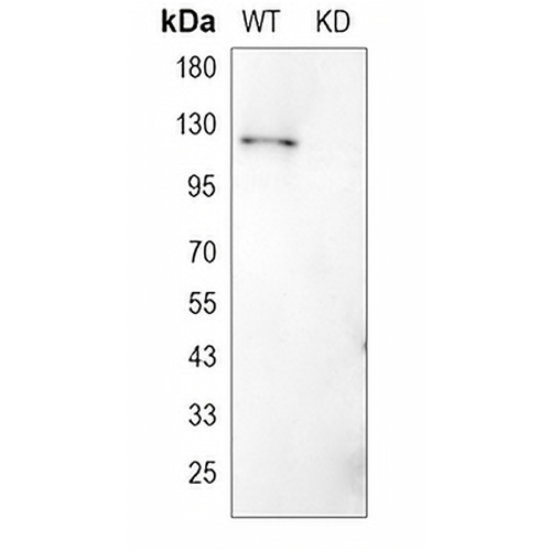
Western blot analysis of Caspase 8 expression in wild type (WT) and knockdown (KD) HeLa cell lysates.
FAQs
 New Products


