CAF-I p60 Rabbit Polyclonal Antibody

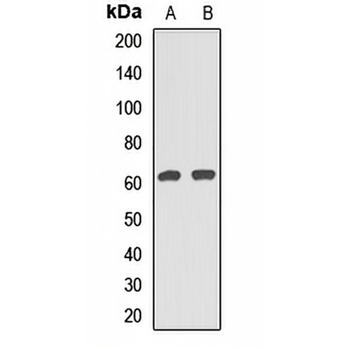
Key features and details

Rabbit polyclonal antibody to CAF-I p60
  • Target: CAF-I p60
  • Source/Host: Rabbit
  • Reactivity: Human
  • Clonality: Polyclonal
  • Applications: WB, IHC, IF/ICC
  • Conjugation: Unconjugated
  • Storage: at-20°C
  • Brand:
CAT.NO. : APA14532
US$ Please choose
US$ Please choose
Size:
Trail, Bulk size or Custom requests Please contact us
Product Details
Background
Acts as a component of the histone chaperone complex chromatin assembly factor 1 (CAF-1), which assembles histone octamers onto DNA during replication and repair. CAF-1 performs the first step of the nucleosome assembly process, bringing newly synthesized histones H3 and H4 to replicating DNA; histones H2A/H2B can bind to this chromatin precursor subsequent to DNA replication to complete the histone octamer.
Application
To ensure optimal assay performance, AREX recommends conducting reagent titration tailored to each testing system for optimal detection results.

WB

1:500 - 1:2000

IHC

1:50 - 1:200

IF/ICC

1:50 - 1:200

*Results are sample-specific. Please refer to your local assay conditions and test parameters for reference.
Overview

Description

Rabbit polyclonal antibody to CAF-I p60

Specificity

Recognizes endogenous levels of CAF-I p60 protein.

Antibody Type

Primary antibody

Imnunogen

Recombinant fusion protein of human CAF-I p60

Purification

The antibody was purified by immunogen affinity chromatography.

Molecular Weight

Predicted: 61 kD; Observed: 61 kD

Form/Buffer

Liquid in 0.42% Potassium phosphate, 0.87% Sodium chloride, pH 7.3, 30% glycerol, and 0.01% sodium azide.

Alternative Names

CAF1A; CAF1P60; MPHOSPH7; MPP7; Chromatin assembly factor 1 subunit B; CAF-1 subunit B; Chromatin assembly factor I p60 subunit; CAF-I 60 kDa subunit; CAF-I p60; M-phase phosphoprotein 7

Gene Symbol

CHAF1B

Entrez Gene

8208(Human)

SwissProt

Q13112(Human)

*AREX continuously optimizes our products. Webpage content may not reflect the latest updates. For inquiries, please contact info@arexbio.com or your local distributor.
*Clone Number, Reactivity, Source/Host and Clonality can be found in the product name and Key Features section above.
Data

Western blot analysis of CAF-I p60 expression in Hela (A), MCF7 (B) whole cell lysates. (Predicted band size: 61 kD; Observed band size: 61 kD)

Immunohistochemical analysis of CAF-I p60 staining in human colon cancer formalin fixed paraffin embedded tissue section. The section was pre-treated using heat mediated antigen retrieval with sodium citrate buffer (pH 6.0). The section was then incubated with the antibody at room temperature and detected using an HRP conjugated compact polymer system. DAB was used as the chromogen. The section was then counterstained with haematoxylin and mounted with DPX.

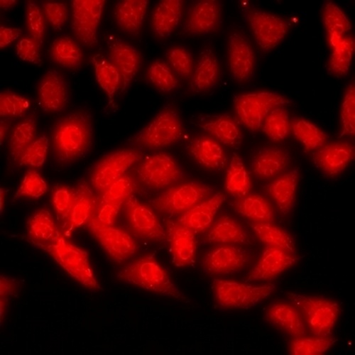
Immunofluorescent analysis of CAF-I p60 staining in MCF7 cells. Formalin-fixed cells were permeabilized with 0.1% Triton X-100 in TBS for 5-10 minutes and blocked with 3% BSA-PBS for 30 minutes at room temperature. Cells were probed with the primary antibody in 3% BSA-PBS and incubated overnight at 4 °C in a humidified chamber. Cells were washed with PBST and incubated with a DyLight 594-conjugated secondary antibody (red) in PBS at room temperature in the dark.

Storage
Store at 4°C short term. For long term storage, store at -20°C, avoiding freeze/thaw cycles.
Note
For Research Use Only. Not for diagnostic, therapeutics, prophylactic or in vivo use.
FAQs
What are the main types of research antibodies and how do they differ?
Research antibodies are mainly divided into monoclonal antibodies and polyclonal antibodies. Monoclonal antibodies typically offer higher specificity and better batch-to-batch consistency, while polyclonal antibodies often provide stronger affinity but may show more variation between batches. The choice depends on your specific experimental needs.
How can I tell if a research antibody is suitable for my experiment?
It is recommended to carefully review the product datasheet for validated applications, species reactivity, recommended dilutions, and published references. For new antibodies, performing a small-scale validation with positive control samples is usually helpful.
Can improper storage of research antibodies affect experimental results?
Yes. Antibodies are sensitive to temperature, repeated freeze-thaw cycles, and contamination. Improper storage may lead to reduced activity, increased background, or weaker signals. It is best to follow the storage instructions provided in the product datasheet.
Why doesn’t the recommended dilution in the datasheet work well in my experiment?
The recommended dilution is based on the supplier’s test conditions. Factors such as sample type, fixation method, and detection system in your lab can influence the optimal working concentration. Performing a dilution series optimization in your own system is often necessary.
What precautions should I take when using a newly purchased research antibody for the first time?
It is advisable to briefly centrifuge the antibody (especially concentrated or lyophilized ones), then perform a small-scale pilot experiment using the recommended conditions. Recording the batch number and usage date is also helpful for future tracking.
New Products
Get in touch with AREX
Name:*
Tel/Phone:*
Company:*
Email:*
Inquiry:
Captcha*
Submitting your email information means that you are willing to receive email information from AREX regarding technology, applications, products, and events. By clicking on the 'unsubscribe' button in the email or by contacting info@arexbio.com You can unsubscribe at any time by sending an email. Regarding your data usage information, please refer to our privacy policy.
© AREX 2024. All Rights Reserved. Sitemap
0.182002s