


BUB1B Rabbit Polyclonal Antibody
 Datasheet
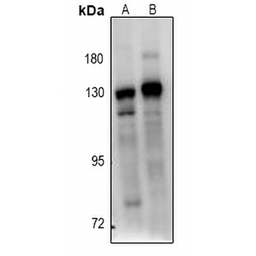
Key features and details
- Target:
- Source/Host:
- Reactivity:
- Clonality:
- Applications:
- Conjugation:
- Storage:
-
Brand:

 Product Details
WB | 1:500 - 1:1000 |
IF/ICC | 1:50 - 1:200 |
Description | Rabbit polyclonal antibody to BUB1B |
Specificity | Recognizes endogenous levels of BUB1B protein. |
Antibody Type | Primary antibody |
Imnunogen | KLH-conjugated synthetic peptide encompassing a sequence within the center region of human BUB1B. The exact sequence is proprietary. |
Purification | The antibody was purified by immunogen affinity chromatography. |
Molecular Weight | Predicted: 119 kD; Observed: 130 kD |
Form/Buffer | Liquid in 0.42% Potassium phosphate, 0.87% Sodium chloride, pH 7.3, 30% glycerol, and 0.01% sodium azide. |
Alternative Names | BUBR1; MAD3L; SSK1; Mitotic checkpoint serine/threonine-protein kinase BUB1 beta; MAD3/BUB1-related protein kinase; hBUBR1; Mitotic checkpoint kinase MAD3L; Protein SSK1 |
Gene Symbol | BUB1B |
Entrez Gene | 701(Human) |
SwissProt | O60566(Human) |
*Clone Number, Reactivity, Source/Host and Clonality can be found in the product name and Key Features section above.
Western blot analysis of BUB1B expression in Hela (A), A549 (B) whole cell lysates. (Predicted band size: 119 kD; Observed band size: 130 kD)
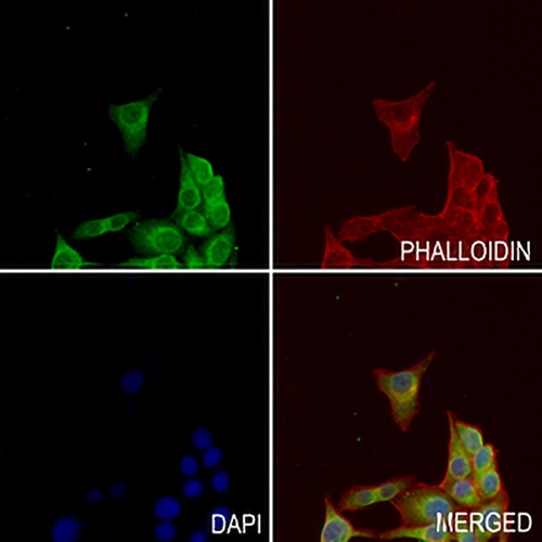
Immunofluorescent analysis of BUB1B staining in MCF7 cells. Formalin-fixed cells were permeabilized with 0.1% Triton X-100 in TBS for 5-10 minutes and blocked with 3% BSA-PBS for 30 minutes at room temperature. Cells were probed with the primary antibody in 3% BSA-PBS and incubated overnight at 4 °C in a hidified chamber. Cells were washed with PBST and incubated with a AREX® Fluor 488 -conjugated secondary antibody (green) in PBS at room temperature in the dark. Phalloidin - AREX® Fluor 594 was used to stain Actin filaments (red). DAPI was used to stain the cell nuclei (blue).
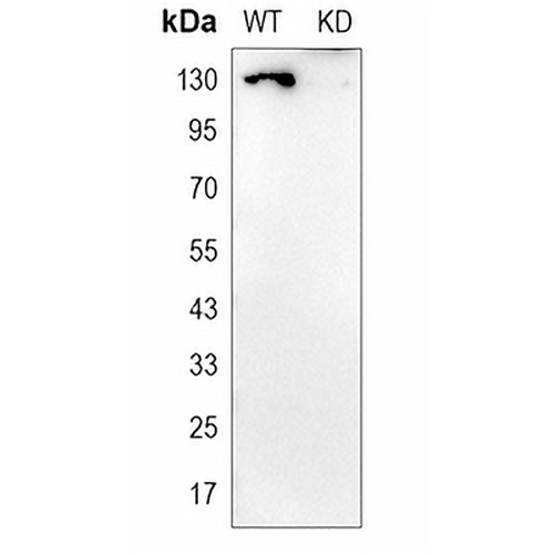
Western blot analysis of BUB1B expression in wild type (WT) and knockdown (KD) HeLa cell lysates.
FAQs
 New Products


