


ATG5 Mouse Monoclonal Antibody(C2533)
 Datasheet
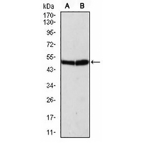
Key features and details
- Target:
- Source/Host:
- Reactivity:
- Clonality:
- Applications:
- Conjugation:
- Storage:
-
Brand:

 Product Details
WB | 1:500 - 1:1000 |
IF/ICC | 1:100 - 1:500 |
FC | 1:100 - 1:200 |
Description | Mouse monoclonal to ATG5 |
Specificity | Recognizes endogenous levels of ATG5 protein |
Antibody Type | Primary antibody |
Imnunogen | Synthesized peptide of human ATG5 |
Purification | This antibody is purified through a protein G column. |
Molecular Weight | Predicted: 32 kD; Observed: 50 kD kD |
Form/Buffer | Mouse IgG2a. Liquid in PBS, pH 7.3, 30% glycerol, and 0.01% sodium azide. |
Alternative Names | APG5L; ASP; Autophagy protein 5; APG5-like; Apoptosis-specifIC, protein |
Gene Symbol | ATG5 |
Entrez Gene | 9474(Human) |
SwissProt | Q9H1Y0(Human) |
*Clone Number, Reactivity, Source/Host and Clonality can be found in the product name and Key Features section above.
Western blot analysis of ATG5 expression in Hela (A), K562 (B) whole cell lysates. (Predicted band size: 32 kD; Observed band size: 50 kD kD)
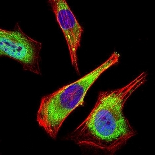
Immunofluorescent analysis of ATG5 staining in Hela cells. Formalin-fixed cells were permeabilized with 0.1% Triton X-100 in TBS for 5-10 minutes and blocked with 3% BSA-PBS for 30 minutes at room temperature. Cells were probed with the primary antibody in 3% BSA-PBS and incubated overnight at 4 °C in a hidified chamber. Cells were washed with PBST and incubated with an AREX® Fluor 488 -conjugated secondary antibody (green) in PBS at room temperature in the dark. Phalloidin - AREX® Fluor 594 was used to stain Actin filaments (red). DAPI was used to stain the cell nuclei (blue).
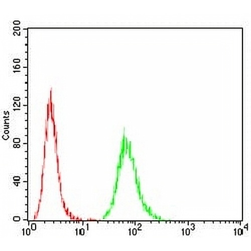
Flow cytometric analysis of HeLa cells using Anti-ATG5 Antibody (green) and negative control (red).
FAQs
 New Products


