


ATG2A Rabbit Polyclonal Antibody
 Datasheet
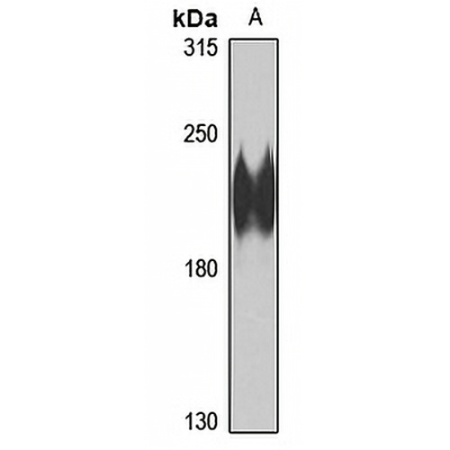
Key features and details
Rabbit polyclonal antibody to ATG2A
- Target:
- Source/Host:
- Reactivity:
- Clonality:
- Applications:
- Conjugation:
- Storage:
-
Brand:

CAT.NO. : APA15120
US$ Please choose
US$ Please choose
 Product Details
Background
Lipid transfer protein involved in autophagosome assembly . Tethers the edge of the isolation membrane (IM) to the endoplasmic reticulum (ER) and mediates direct lipid transfer from ER to IM for IM expansion . Binds to the ER exit site (ERES), which is the membrane source for autophagosome formation, and extracts phospholipids from the membrane source and transfers them to ATG9 (ATG9A or ATG9B) to the IM for membrane expansion . Lipid transfer activity is enhanced by WIPI1 and WDR45/WIPI4, which promote ATG2A-association with phosphatidylinositol 3-monophosphate (PI3P)-containing membranes . Also regulates lipid droplets morphology and distribution within the cell .
Application
To ensure optimal assay performance, AREX recommends conducting reagent titration tailored to each testing system for optimal detection results.
*Results are sample-specific. Please refer to your local assay conditions and test parameters for reference.
WB | 1:500 - 1:2000 |
Overview
Description | Rabbit polyclonal antibody to ATG2A |
Specificity | Recognizes endogenous levels of ATG2A protein |
Antibody Type | Primary antibody |
Imnunogen | KLH-conjugated synthetic peptide of human ATG2A. The exact sequence is proprietary. |
Purification | The antibody was purified by immunogen affinity chromatography. |
Molecular Weight | Predicted: 13; Observed: 213 kD |
Form/Buffer | Liquid in 0.42% Potassium phosphate, 0.87% Sodium chloride, pH 7.3, 30% glycerol, and 0.01% sodium azide. |
Alternative Names | KIAA0404; Autophagy-related protein 2 homolog A |
Gene Symbol | ATG2A |
Entrez Gene | 23130(Human) |
SwissProt | Q2TAZ0(Human) |
*Clone Number, Reactivity, Source/Host and Clonality can be found in the product name and Key Features section above.
Data
Western blot analysis of ATG2A expression in HeLa (A) whole cell lysates. (Predicted band size: 13; 35; 212; 213 kD; Observed band size: 213 kD)
Storage
Store at 4°C short term. For long term storage, store at -20°C, avoiding freeze/thaw cycles.
Note
For Research Use Only. Not for diagnostic, therapeutics, prophylactic or in vivo use.
FAQs
What are the main types of research antibodies and how do they differ?

Research antibodies are mainly divided into monoclonal antibodies and polyclonal antibodies. Monoclonal antibodies typically offer higher specificity and better batch-to-batch consistency, while polyclonal antibodies often provide stronger affinity but may show more variation between batches. The choice depends on your specific experimental needs.
How can I tell if a research antibody is suitable for my experiment?
It is recommended to carefully review the product datasheet for validated applications, species reactivity, recommended dilutions, and published references. For new antibodies, performing a small-scale validation with positive control samples is usually helpful.
Can improper storage of research antibodies affect experimental results?
Yes. Antibodies are sensitive to temperature, repeated freeze-thaw cycles, and contamination. Improper storage may lead to reduced activity, increased background, or weaker signals. It is best to follow the storage instructions provided in the product datasheet.
Why doesn’t the recommended dilution in the datasheet work well in my experiment?
The recommended dilution is based on the supplier’s test conditions. Factors such as sample type, fixation method, and detection system in your lab can influence the optimal working concentration. Performing a dilution series optimization in your own system is often necessary.
What precautions should I take when using a newly purchased research antibody for the first time?
It is advisable to briefly centrifuge the antibody (especially concentrated or lyophilized ones), then perform a small-scale pilot experiment using the recommended conditions. Recording the batch number and usage date is also helpful for future tracking.
New Products


