


Alpha smooth muscle Actin Rabbit Monoclonal Antibody(ARA756)
 Datasheet
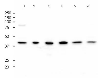
Key features and details
- Target:
- Source/Host:
- Reactivity:
- Clonality:
- Applications:
- Conjugation:
- Storage:
-
Brand:

 Product Details
WB | 1:1000-1:5000 |
IF/ICC | 1:2000-1:10000 |
FC | 1:200-1:1000 |
Description | Rabbit Monoclonal Antibody to Alpha smooth muscle Actin |
Antibody Type | Primary antibody |
Predicted MW | 42kDa |
Immunogen | A synthetic peptide corresponding to residues on the N-terminus of human alpha smooth muscle actin was used as an immunogen. |
Purification | ProA affinity purified IgG |
Form/Buffer | PBS 59%, Sodium azide 0.01%, Glycerol 40%, BSA 0.31%. |
Alternative Names | ACTA2; ACTSA; ACTVS; GIG46; Actin aortic smooth muscle; Alpha-actin-2; Cell growth-inhibiting gene 46 protein |
Gene Symbol | ACTA2 |
Entrez Gene | 59(Human); 11475(Mouse); 81633(Rat) |
Swissprot | P62736 |
*Clone Number, Reactivity, Source/Host and Clonality can be found in the product name and Key Features section above.
All lanes: Anti-alpha smooth muscle Actin antibody at 1:5,000 dilution
Predicted MW: 42 kDa
Observed MW: 42 kDa
Lane 1: Rat Testis
Lane 2: A549
Lane 3: Rat Brain
Lane 4: Hela
Lane 5: HepG2
Lane 6: K562
Lysates at 10 µg per lane
2nd Ab:
G&R HRP(H+L) 1:10,000
Exposure: 100s
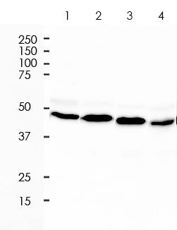
All lanes: Anti-alpha smooth muscle Actin antibody at 1:5,000 dilution
Predicted MW: 42 kDa
Observed MW: 42 kDa
Lane 1: Mu Brain
Lane 2: Mu Heart
Lane 3: Rat Kidney
Lane 4: Rat Liver
Lysates at 10 µg per lane
2nd Ab:
G&R HRP(H+L) 1:10,000
Exposure: 20s
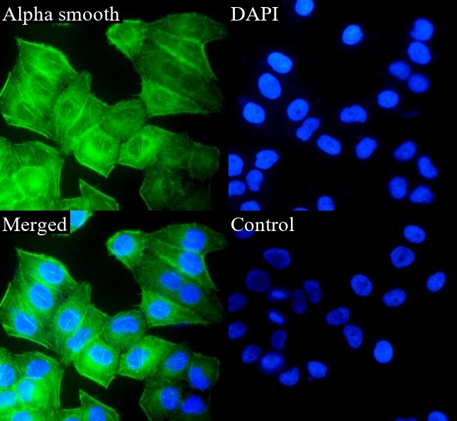
ARA756 staining Alpha smooth muscle Actin in Hela cells by IF/ICC (immunofluorescence/immunocytochemistry). Cells were fixed with paraformaldehyde, permeabilized with 0.1% Triton X-100 and blocked with 10% goat serum for half an hour at room temperature. Samples were incubated with primary antibody (1:10,000) at 4°C. An Alexa Fluor<sup>®</sup> 488-conjugated Goat Anti-Rabbit IgG polyclonal was used as the secondary antibody (1:500). DAPI (blue) was used as the nuclear counter stain.
Control: PBS and secondary antibody, An Alexa Fluor<sup>®</sup> 488-conjugated Goat Anti-Rabbit IgG (1:500).
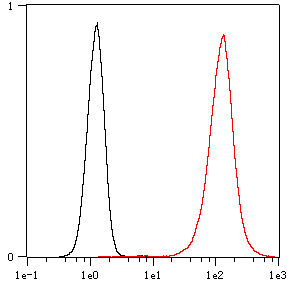
Overlay histogram showing Hela cells stained with ARA756 (Red). The cells were fixed with 4% paraformaldehyde (10 min) and then permeabilized with 0.1% TritonX-100 for 15 min. The cells were then incubated in the antibody (ARA756, 1:1,000 dilution) in 1x PBS/1% BSA for 30 min at room temperature. The secondary antibody used was a Goat Anti-Rabbit Alexa Fluor<sup>®</sup> 488 (IgG H+L) at 1:2,000 dilution for 20 min at room temperature. Unlabelled sample (Black) was used as a control.
FAQs
 New Products


