


SUMO1 Mouse Monoclonal Antibody(C3852)
 Datasheet
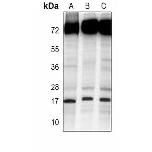
Key features and details
- Target:
- Source/Host:
- Reactivity:
- Clonality:
- Applications:
- Conjugation:
- Storage:
-
Brand:

 Product Details
WB | 1:500 - 1:2000 |
IHC | 1:50 - 1:200 |
IF/ICC | 1:10 - 1:50 |
FC | 1:10 - 1:50 |
Description | Mouse monoclonal antibody to SUMO1 |
Specificity | Recognizes endogenous levels of SUMO1 protein. |
Antibody Type | Primary antibody |
Imnunogen | Recombinant fusion protein of human SUMO1. The exact sequence is proprietary. |
Purification | This antibody is purified through a protein G column. |
Molecular Weight | Predicted: 11 kD; Observed: 17; 72 kD |
Form/Buffer | Mouse IgG1. Liquid in PBS, pH 7.3, 30% glycerol, and 0.01% sodium azide. |
Alternative Names | SMT3C; SMT3H3; UBL1; Small ubiquitin-related modifier 1; SUMO-1; GAP-modifying protein 1; GMP1; SMT3 homolog 3; Sentrin; Ubiquitin-homology domain protein PIC1; Ubiquitin-like protein SMT3C; Smt3C; Ubiquitin-like protein UBL1 |
Gene Symbol | SUMO1 |
Entrez Gene | 7341(Human) |
SwissProt | P63165(Human) |
*Clone Number, Reactivity, Source/Host and Clonality can be found in the product name and Key Features section above.
Western blot analysis of SUMO1 expression in HL60 (A), Hela (B), Jurkat (C) whole cell lysates. (Predicted band size: 11 kD; Observed band size: 17; 72 kD)
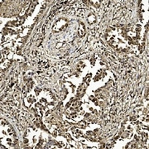
Immunohistochemical analysis of SUMO1 staining in human lung adenocarcinoma formalin fixed paraffin embedded tissue section. The section was pre-treated using heat mediated antigen retrieval with sodium citrate buffer (pH 6.0). The section was then incubated with the antibody at room temperature and detected using an HRP conjugated compact polymer system. DAB was used as the chromogen. The section was then counterstained with haematoxylin and mounted with DPX.
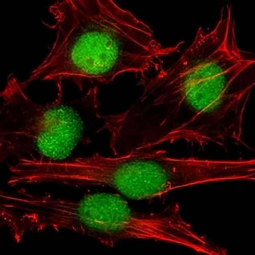
Immunofluorescent analysis of SUMO1 staining in Hela cells. Formalin-fixed cells were permeabilized with 0.1% Triton X-100 in TBS for 5-10 minutes and blocked with 3% BSA-PBS for 30 minutes at room temperature. Cells were probed with the primary antibody in 3% BSA-PBS and incubated overnight at 4 °C in a humidified chamber. Cells were washed with PBST and incubated with a AREX® Fluor 488 -conjugated secondary antibody (green) in PBS at room temperature in the dark. Phalloidin - AREX® Fluor 555 was used to stain Actin filaments (red). DAPI was used to stain the cell nuclei (blue).
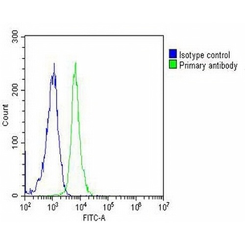
Flow cytometric analysis of Jurkat cells using Anti-SUMO1 Antibody. The cells were fixed with 2% paraformaldehyde (10 min) and then permeabilized with 90% methanol for 10 min. The cells were incubated in 2% bovine serum albumin to block non-specific protein-protein interactions followed by the antibody at 37 °C for 60 min. The secondary antibody Goat Anti-Mouse IgG (H&L) - AREX® Fluor 488 was incubated at 37 °C for 40 min. Isotype control antibody (blue line) was used under the same condition.
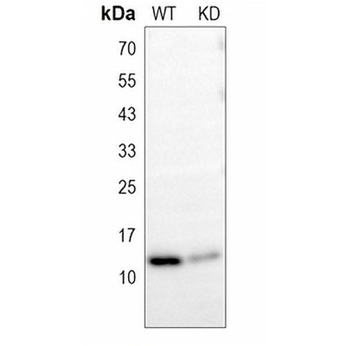
Western blot analysis of SUMO1 expression in wild type (WT) and knockdown (KD) HeLa cell lysates.
FAQs
 New Products


