PGK1 Mouse Monoclonal Antibody(C3554)

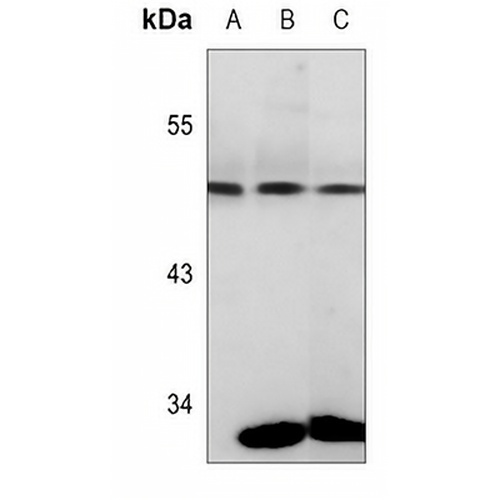
Key features and details

Mouse monoclonal antibody to PGK1
  • Target: PGK1
  • Source/Host: Mouse
  • Reactivity: Human, Mouse
  • Clonality: Monoclonal
  • Applications: WB, IHC, FC
  • Conjugation: Unconjugated
  • Storage: at-20°C
  • Brand:
CAT.NO. : AMA03166
US$ Please choose
US$ Please choose
Size:
Trail, Bulk size or Custom requests Please contact us
Product Details
Background
Catalyzes one of the two ATP producing reactions in the glycolytic pathway via the reversible conversion of 1,3-diphosphoglycerate to 3-phosphoglycerate . Both L- and D- forms of purine and pyrimidine nucleotides can be used as substrates, but the activity is much lower on pyrimidines . In addition to its role as a glycolytic enzyme, it seems that PGK1 acts as a polymerase alpha cofactor protein (primer recognition protein) . Acts as a protein kinase when localized to the mitochondrion where it phosphorylates pyruvate dehydrogenase kinase PDK1 to inhibit pyruvate dehydrogenase complex activity and suppress the formation of acetyl-coenzyme A from pyruvate, and consequently inhibit oxidative phosphorylation and promote glycolysis . May play a role in sperm motility .
Application
To ensure optimal assay performance, AREX recommends conducting reagent titration tailored to each testing system for optimal detection results.

WB

1:500 - 1:2000

IHC

1:50 - 1:100

FC

1:50 - 1:100

*Results are sample-specific. Please refer to your local assay conditions and test parameters for reference.
Overview

Description

Mouse monoclonal antibody to PGK1

Specificity

Recognizes endogenous levels of PGK1 protein.

Antibody Type

Primary antibody

Imnunogen

KLH-conjugated synthetic peptide encompassing a sequence within the center region of human PGK1. The exact sequence is proprietary.

Purification

The antibody was purified by immunogen affinity chromatography.

Molecular Weight

Predicted: 44 kD; Observed: 44 kD

Form/Buffer

Mouse IgG2a kappa. Liquid in PBS, pH 7.3, 30% glycerol, and 0.01% sodium azide.

Alternative Names

PGKA; Phosphoglycerate kinase 1; Cell migration-inducing gene 10 protein; Primer recognition protein 2; PRP 2

Gene Symbol

PGK1

Entrez Gene

5230(Human); 18655(Mouse)

SwissProt

P00558(Human); P09411(Mouse)

*AREX continuously optimizes our products. Webpage content may not reflect the latest updates. For inquiries, please contact info@arexbio.com or your local distributor.
*Clone Number, Reactivity, Source/Host and Clonality can be found in the product name and Key Features section above.
Data

Western blot analysis of PGK1 expression in HEK293T (A), A431 (B), mouse brain (C) whole cell lysates. (Predicted band size: 44 kD; Observed band size: 44 kD)

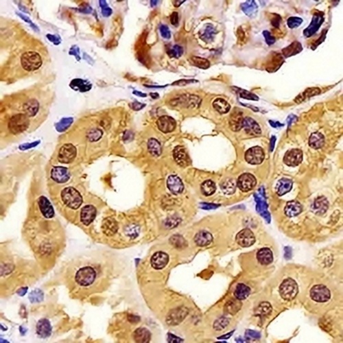
Immunohistochemical analysis of PGK1 staining in human kidney formalin fixed paraffin embedded tissue section. The section was pre-treated using heat mediated antigen retrieval with sodium citrate buffer (pH 6.0). The section was then incubated with the antibody at room temperature and detected using an HRP conjugated compact polymer system. DAB was used as the chromogen. The section was then counterstained with haematoxylin and mounted with DPX.

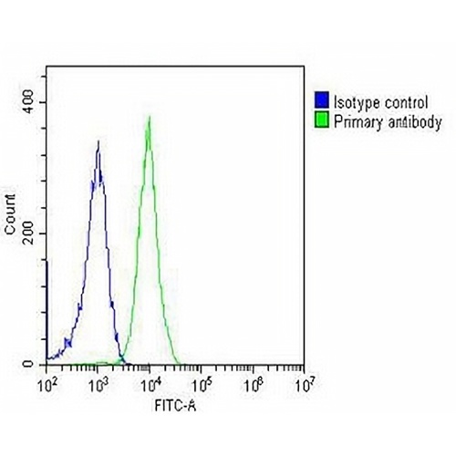
Overlay histogram showing Jurkat cells stained with Anti-PGK1 Antibody (green line). The cells were fixed with 2% paraformaldehyde (10 min) and then permeabilized with 90% methanol for 10 min. The cells were then icubated in 2% bovine serum albumin to block non-specific protein-protein interactions followed by Anti-PGK1 Antibody for 60 min at 37 °C. The secondary antibody used was Goat Anti-Mouse IgG (H&L) - AREX® Fluor 488 at 1/200 dilution for 40 min at 37 °C. Isotype control antibody (blue line) was mouse IgG2a (1μg/1x10^6 cells) used under the same conditions. Acquisition of >10, 000 events was performed.

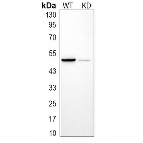
Western blot analysis of PGK1 expression in wild type (WT) and knockdown (KD) HeLa cell lysates.

Storage
Store at 4°C short term. For long term storage, store at -20°C, avoiding freeze/thaw cycles.
Note
For Research Use Only. Not for use in diagnostic procedures.
FAQs
What are the main types of research antibodies and how do they differ?
Research antibodies are mainly divided into monoclonal antibodies and polyclonal antibodies. Monoclonal antibodies typically offer higher specificity and better batch-to-batch consistency, while polyclonal antibodies often provide stronger affinity but may show more variation between batches. The choice depends on your specific experimental needs.
How can I tell if a research antibody is suitable for my experiment?
It is recommended to carefully review the product datasheet for validated applications, species reactivity, recommended dilutions, and published references. For new antibodies, performing a small-scale validation with positive control samples is usually helpful.
Can improper storage of research antibodies affect experimental results?
Yes. Antibodies are sensitive to temperature, repeated freeze-thaw cycles, and contamination. Improper storage may lead to reduced activity, increased background, or weaker signals. It is best to follow the storage instructions provided in the product datasheet.
Why doesn’t the recommended dilution in the datasheet work well in my experiment?
The recommended dilution is based on the supplier’s test conditions. Factors such as sample type, fixation method, and detection system in your lab can influence the optimal working concentration. Performing a dilution series optimization in your own system is often necessary.
What precautions should I take when using a newly purchased research antibody for the first time?
It is advisable to briefly centrifuge the antibody (especially concentrated or lyophilized ones), then perform a small-scale pilot experiment using the recommended conditions. Recording the batch number and usage date is also helpful for future tracking.
New Products
Get in touch with AREX
Name:*
Tel/Phone:*
Company:*
Email:*
Inquiry:
Captcha*
Submitting your email information means that you are willing to receive email information from AREX regarding technology, applications, products, and events. By clicking on the 'unsubscribe' button in the email or by contacting info@arexbio.com You can unsubscribe at any time by sending an email. Regarding your data usage information, please refer to our privacy policy.
© AREX 2024. All Rights Reserved. Sitemap
0.597316s