


PGD Rabbit Monoclonal Antibody(C3070)
 Datasheet
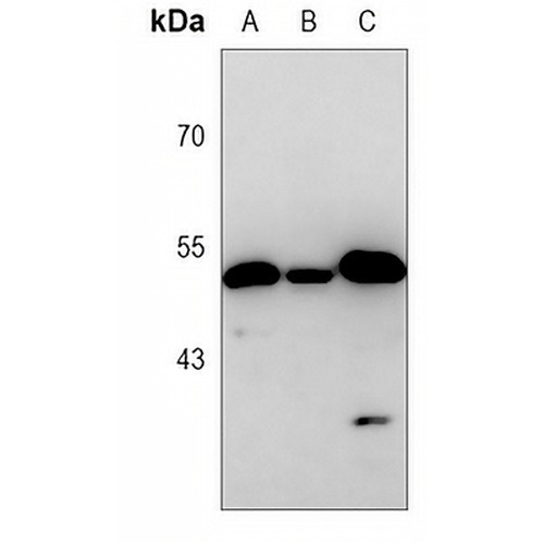
Key features and details
- Target:
- Source/Host:
- Reactivity:
- Clonality:
- Applications:
- Conjugation:
- Storage:
-
Brand:

 Product Details
WB | 1:500 - 1:1000 |
IF/ICC | 1:50 - 1:100 |
IP | 1:10 - 1:50 |
Description | Rabbit monoclonal antibody to PGD |
Specificity | Recognizes endogenous levels of PGD protein. |
Antibody Type | Primary antibody |
Imnunogen | A synthetic peptide of human PGD |
Purification | The antibody was purified by immunogen affinity chromatography. |
Molecular Weight | Predicted: 53 kD; Observed: 49 kD |
Form/Buffer | Liquid in 50mM Tris-Glycine (pH 7.4), 0.15M NaCl, 50% Glycerol, 0.01% Sodium azide and 0.05% BSA. |
Alternative Names | PGDH; 6-phosphogluconate dehydrogenase decarboxylating |
Gene Symbol | PGD |
Entrez Gene | 5226(Human); 110208(Mouse); 100360180(Rat) |
SwissProt | P52209(Human); Q9DCD0(Mouse); P85968(Rat) |
*Clone Number, Reactivity, Source/Host and Clonality can be found in the product name and Key Features section above.
Western blot analysis of PGD expression in C6 (A), NIH3T3 (B), Hela (C) whole cell lysates. (Predicted band size: 53 kD; Observed band size: 49 kD)
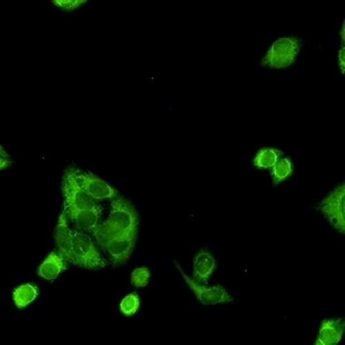
Immunofluorescent analysis of PGD staining in HeLa cells. Formalin-fixed cells were permeabilized with 0.1% Triton X-100 in TBS for 5-10 minutes and blocked with 3% BSA-PBS for 30 minutes at room temperature. Cells were probed with the primary antibody in 3% BSA-PBS and incubated overnight at 4 °C in a hidified chamber. Cells were washed with PBST and incubated with a AREX® Fluor 488 -conjugated secondary antibody (green) in PBS at room temperature in the dark.
FAQs
 New Products


