


Peroxiredoxin 5 Rabbit Polyclonal Antibody
 Datasheet
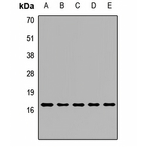
Key features and details
- Target:
- Source/Host:
- Reactivity:
- Clonality:
- Applications:
- Conjugation:
- Storage:
-
Brand:

 Product Details
WB | 1:500 - 1:1000 |
IF/ICC | 1:50 - 1:200 |
Description | Rabbit polyclonal antibody to Peroxiredoxin 5 |
Specificity | Recognizes endogenous levels of Peroxiredoxin 5 protein. |
Antibody Type | Primary antibody |
Imnunogen | Recombinant fusion protein of human Peroxiredoxin 5 |
Purification | The antibody was purified by immunogen affinity chromatography. |
Molecular Weight | Predicted: 12; Observed: 17 kD |
Form/Buffer | Liquid in 0.42% Potassium phosphate, 0.87% Sodium chloride, pH 7.3, 30% glycerol, and 0.01% sodium azide. |
Alternative Names | ACR1; Peroxiredoxin-5 mitochondrial; Alu corepressor 1; Antioxidant enzyme B166; AOEB166; Liver tissue 2D-page spot 71B; PLP; Peroxiredoxin V; Prx-V; Peroxisomal antioxidant enzyme; TPx type VI; Thioredoxin peroxidase PMP20; Thioredoxin reductase |
Gene Symbol | PRDX5 |
Entrez Gene | 25824(Human); 54683(Mouse); 113898(Rat) |
SwissProt | P30044(Human); P99029(Mouse); Q9R063(Rat) |
*Clone Number, Reactivity, Source/Host and Clonality can be found in the product name and Key Features section above.
Western blot analysis of Peroxiredoxin 5 expression in Hela (A), HepG2 (B), mouse liver (C), mouse heart (D), rat brain (E) whole cell lysates. (Predicted band size: 12; 17; 22 kD; Observed band size: 17 kD)

Immunofluorescent analysis of Peroxiredoxin 5 staining in U2OS cells. Formalin-fixed cells were permeabilized with 0.1% Triton X-100 in TBS for 5-10 minutes and blocked with 3% BSA-PBS for 30 minutes at room temperature. Cells were probed with the primary antibody in 3% BSA-PBS and incubated overnight at 4 °C in a humidified chamber. Cells were washed with PBST and incubated with a DyLight 594-conjugated secondary antibody (red) in PBS at room temperature in the dark.
FAQs
 New Products


