


p38 (Phospho-T180) Rabbit Polyclonal Antibody
 Datasheet
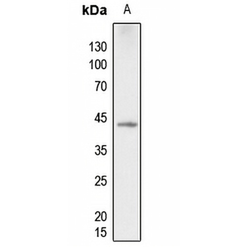
Key features and details
- Target:
- Source/Host:
- Reactivity:
- Clonality:
- Applications:
- Conjugation:
- Storage:
-
Brand:

 Product Details
WB | 1:500 - 1:1000 |
IF/ICC | 1:50 - 1:200 |
Description | Rabbit polyclonal antibody to p38 (Phospho-T180) |
Specificity | Recognizes endogenous levels of p38 protein only when phosphorylated at T180. |
Antibody Type | Primary antibody |
Imnunogen | KLH-conjugated synthetic phosphopeptide corresponding to residues surrounding T180 of human p38 protein. The exact sequence is proprietary. |
Purification | The antibody was purified by immunogen affinity chromatography. |
Molecular Weight | Predicted: 41 kD; Observed: 43 kD |
Form/Buffer | Liquid in 0.42% Potassium phosphate, 0.87% Sodium chloride, pH 7.3, 30% glycerol, and 0.01% sodium azide. |
Alternative Names | CSBP; CSBP1; CSBP2; CSPB1; MXI2; SAPK2A; Mitogen-activated protein kinase 14; MAP kinase 14; MAPK 14; Cytokine suppressive anti-inflammatory drug-binding protein; CSAID-binding protein; CSBP; MAP kinase MXI2; MAX-interacting protein 2; Mitogen-activated protein kinase p38 alpha; MAP kinase p38 alpha; Stress-activated protein kinase 2a; SAPK2a |
Gene Symbol | MAPK14 |
Entrez Gene | 1432(Human); 26416(Mouse) |
SwissProt | Q16539(Human); P47811(Mouse); P70618(Rat) |
*Clone Number, Reactivity, Source/Host and Clonality can be found in the product name and Key Features section above.
Western blot analysis of p38 (Phospho-T180) expression in zebrafish (A) whole cell lysates. (Predicted band size: 41 kD; Observed band size: 43 kD)
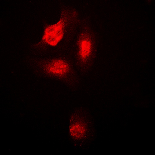
Immunofluorescent analysis of p38 (Phospho-T180) staining in HepG2 cells. Formalin-fixed cells were permeabilized with 0.1% Triton X-100 in TBS for 5-10 minutes and blocked with 3% BSA-PBS for 30 minutes at room temperature. Cells were probed with the primary antibody in 3% BSA-PBS and incubated overnight at 4 °C in a humidified chamber. Cells were washed with PBST and incubated with a DyLight 594-conjugated secondary antibody (red) in PBS at room temperature in the dark.
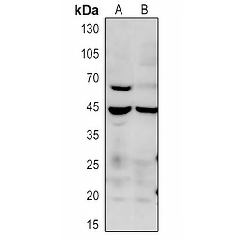
Western blot analysis of p38 (Phospho-T180) expression in mouse liver (A), rat liver (B) whole cell lysates.
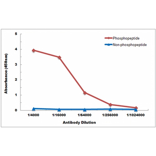
Direct ELISA antibody dose-response curve using Anti-p38 (Phospho-T180) Antibody. Antigen (Phosphopeptide and non-phosphopeptide) concentration is 5 ug/ml. Goat Anti-Rabbit IgG (H&L) - HRP was used as the secondary antibody, and signal was developed by TMB substrate.
FAQs
 New Products


