


MARK3 Mouse Monoclonal Antibody(C2832)
 Datasheet
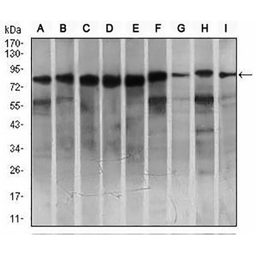
Key features and details
- Target:
- Source/Host:
- Reactivity:
- Clonality:
- Applications:
- Conjugation:
- Storage:
-
Brand:

 Product Details
WB | 1:500 - 1:1000 |
IHC | 1:100 - 1:500 |
FC | 1:100 - 1:200 |
Description | Mouse monoclonal to MARK3 |
Specificity | Recognizes endogenous levels of MARK3 protein |
Antibody Type | Primary antibody |
Imnunogen | Recombinant fusion protein of human MARK3 expressed in E. Coli |
Purification | This antibody is purified through a protein G column. |
Molecular Weight | Predicted: 87 kD; Observed: 87 kD kD |
Form/Buffer | Mouse IgG1. Liquid in PBS, pH 7.3, 30% glycerol, and 0.01% sodium azide. |
Alternative Names | CTAK1; EMK2; MAP/mIC,rotubule affinity-regulating kinase 3; C-TAK1; cTAK1; Cdc25C-associated protein kinase 1; ELKL motif kinase 2; EMK-2; Protein kinase STK10; Ser/Thr protein kinase PAR-1; Par-1a; Serine/threonine-protein kinase p78 |
Gene Symbol | MARK3 |
Entrez Gene | 4140(Human); 170577(Rat) |
SwissProt | P27448(Human); Q03141(Mouse) |
*Clone Number, Reactivity, Source/Host and Clonality can be found in the product name and Key Features section above.
Western blot analysis of MARK3 expression in Hela (A), SKNSH (B), K562 (C), HCT116 (D), HEK293 (E), 3T3L1 (F), NIH3T3 (G), Jurkat (H), A431 (I) whole cell lysates. (Predicted band size: 87 kD; Observed band size: 87 kD kD)
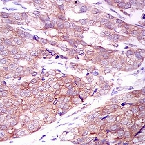
Immunohistochemical analysis of MARK3 staining in human bladder cancer formalin fixed paraffin embedded tissue section. The section was pre-treated using heat mediated antigen retrieval with sodium citrate buffer (pH 6.0). The section was then incubated with the antibody at room temperature and detected using an HRP conjugated compact polymer system. DAB was used as the chromogen. The section was then counterstained with haematoxylin and mounted with DPX.
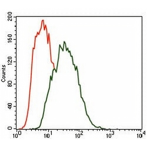
Flow cytometric analysis of SKNSH cells using Anti-MARK3 Antibody (green) and negative control (red).
FAQs
 New Products


