


ILF3 Rabbit Monoclonal Antibody(C3164)
 Datasheet
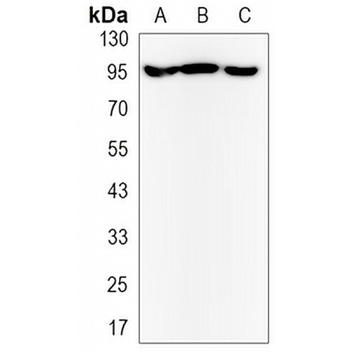
Key features and details
- Target:
- Source/Host:
- Reactivity:
- Clonality:
- Applications:
- Conjugation:
- Storage:
-
Brand:

 Product Details
WB | 1:500 - 1:1000 |
IHC | 1:50 - 1:100 |
IF/ICC | 1:50 - 1:100 |
IP | 1:10 - 1:50 |
Description | Rabbit monoclonal antibody to ILF3 |
Specificity | Recognizes endogenous levels of ILF3 protein. |
Antibody Type | Primary antibody |
Imnunogen | A synthetic peptide of human ILF3 |
Purification | The antibody was purified by immunogen affinity chromatography. |
Molecular Weight | Predicted: 95 kD; Observed: 95 kD |
Form/Buffer | Liquid in 50mM Tris-Glycine (pH 7.4), 0.15M NaCl, 50% Glycerol, 0.01% Sodium azide and 0.05% BSA. |
Alternative Names | DRBF; MPHOSPH4; NF90; Interleukin enhancer-binding factor 3; Double-stranded RNA-binding protein 76; DRBP76; M-phase phosphoprotein 4; MPP4; Nuclear factor associated with dsRNA; NFAR; Nuclear factor of activated T-cells 90 kDa; NF-AT-90; Translational control protein 80; TCP80 |
Gene Symbol | ILF3 |
Entrez Gene | 3609(Human); 84472(Rat) |
SwissProt | Q12906(Human); Q9JIL3(Rat) |
*Clone Number, Reactivity, Source/Host and Clonality can be found in the product name and Key Features section above.
Western blot analysis of ILF3 expression in Jurkat (A), C6 (B), Hela (C) whole cell lysates. (Predicted band size: 95 kD; Observed band size: 95 kD)
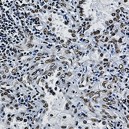
Immunohistochemical analysis of ILF3 staining in human lung cancer formalin fixed paraffin embedded tissue section. The section was pre-treated using heat mediated antigen retrieval with sodium citrate buffer (pH 6.0). The section was then incubated with the antibody at room temperature and detected using an HRP conjugated compact polymer system. DAB was used as the chromogen. The section was then counterstained with haematoxylin and mounted with DPX.
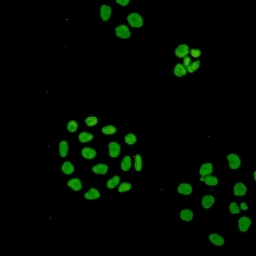
Immunofluorescent analysis of ILF3 staining in HeLa cells. Formalin-fixed cells were permeabilized with 0.1% Triton X-100 in TBS for 5-10 minutes and blocked with 3% BSA-PBS for 30 minutes at room temperature. Cells were probed with the primary antibody in 3% BSA-PBS and incubated overnight at 4 °C in a hidified chamber. Cells were washed with PBST and incubated with a AREX® Fluor 488 -conjugated secondary antibody (green) in PBS at room temperature in the dark.
FAQs
 New Products


