


Histone H4 (MonoMethyl-K12) Rabbit Polyclonal Antibody
 Datasheet
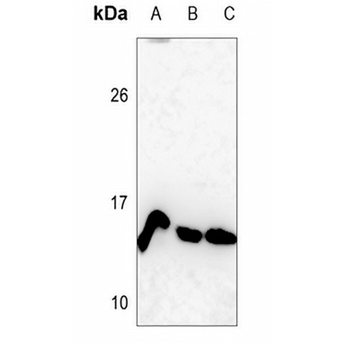
Key features and details
- Target:
- Source/Host:
- Reactivity:
- Clonality:
- Applications:
- Conjugation:
- Storage:
-
Brand:

 Product Details
WB | 1:500 - 1:1000 |
IF/ICC | 1:50 - 1:200 |
Description | Rabbit polyclonal antibody to Histone H4 (MonoMethyl-K12) |
Specificity | Recognizes endogenous levels of Histone H4 protein only when Mono-methylated at K12. |
Antibody Type | Primary antibody |
Imnunogen | KLH-conjugated synthetic Mono-methylated peptide corresponding to residues surrounding K12 of human Histone H4 protein. The exact sequence is proprietary. |
Purification | The antibody was purified by immunogen affinity chromatography. |
Molecular Weight | Predicted: 11 kD; Observed: 14 kD |
Form/Buffer | Liquid in 0.42% Potassium phosphate, 0.87% Sodium chloride, pH 7.3, 30% glycerol, and 0.01% sodium azide. |
Alternative Names | H4/A; H4FA; H4/I; H4FI; H4/G; H4FG; H4/B; H4FB; H4/J; H4FJ; H4/C; H4FC; H4/H; H4FH; H4/M; H4FM; H4/E; H4FE; H4/D; H4FD; H4/K; H4FK; H4/N; H4F2; H4FN; HIST2H4; H4/O; H4FO; Histone H4 |
Gene Symbol | HIST1H4A; HIST1H4B; HIST1H4C; HIST1H4D; HIST1H4E; HIST1H4F; HIST1H4H; HIST1H4I; HIST1H4J; HIST1H4K; HIST1H4L; HIST2H4A; HIST2H4B; HIST4H4 |
Entrez Gene | 121504; 554313; 8294; 8359; 8360; 8361; 8362; 8363; 8364; 8365; 8366; 8367; 8368; 8370(Human); 100041230; 102641229; 319155; 319156; 319157; 319158; 319159; 319160; 319161; 320332; 326619; 326620; 69386; 97122(Mouse); 100360950; 100912290; 100912418; 100912564; 102548682; 102551184; 102557184; 291152; 295277; 500351; 502913; 64627; 680097(Rat) |
SwissProt | P62805(Human); P62806(Mouse); P62804(Rat) |
*Clone Number, Reactivity, Source/Host and Clonality can be found in the product name and Key Features section above.
Western blot analysis of Histone H4 (MonoMethyl-K12) expression in HEK293T (A), H446 (B), H1688 (C) whole cell lysates. (Predicted band size: 11 kD; Observed band size: 14 kD)
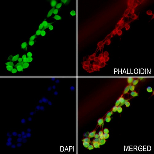
Immunofluorescent analysis of Histone H4 (MonoMethyl-K12) staining in C6 cells. Formalin-fixed cells were permeabilized with 0.1% Triton X-100 in TBS for 5-10 minutes and blocked with 3% BSA-PBS for 30 minutes at room temperature. Cells were probed with the primary antibody in 3% BSA-PBS and incubated overnight at 4 °C in a hidified chamber. Cells were washed with PBST and incubated with a AREX® Fluor 488 -conjugated secondary antibody (green) in PBS at room temperature in the dark. Phalloidin - AREX® Fluor 594 was used to stain Actin filaments (red). DAPI was used to stain the cell nuclei (blue).
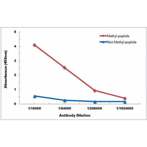
Direct ELISA antibody dose-response curve using Anti-Histone H4 (MonoMethyl-K12) Antibody. Antigen (methyl-peptide and non-methyl-peptide) concentration is 5 ug/ml. Goat Anti-Rabbit IgG (H&L) - HRP was used as the secondary antibody, and signal was developed by TMB substrate.
FAQs
 New Products


