


Histone H3 (Butyryl-K23) Rabbit Polyclonal Antibody
 Datasheet
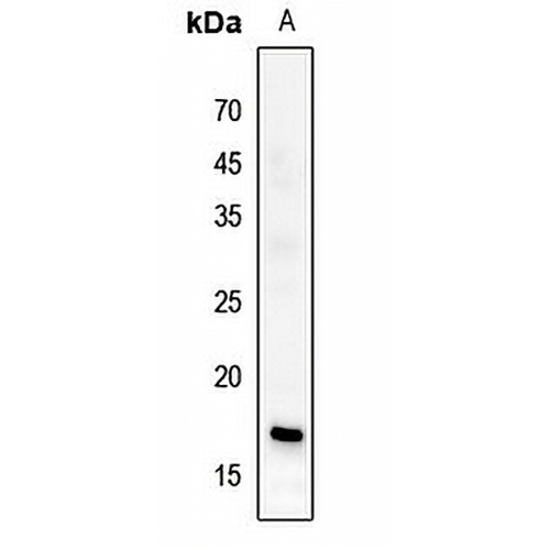
Key features and details
- Target:
- Source/Host:
- Reactivity:
- Clonality:
- Applications:
- Conjugation:
- Storage:
-
Brand:

 Product Details
WB | 1:500 - 1:1000 |
IF/ICC | 1:50 - 1:200 |
Description | Rabbit polyclonal antibody to Histone H3 (Butyryl-K23) |
Specificity | Recognizes endogenous levels of Histone H3 protein only when butylated at K23. |
Antibody Type | Primary antibody |
Imnunogen | KLH-conjugated synthetic butylated peptide corresponding to residues surrounding K23 of human Histone H3 protein. The exact sequence is proprietary. |
Purification | The antibody was purified by immunogen affinity chromatography. |
Molecular Weight | Predicted: 15 kD; Observed: 17 kD |
Form/Buffer | Liquid in 0.42% Potassium phosphate, 0.87% Sodium chloride, pH 7.3, 30% glycerol, and 0.01% sodium azide. |
Alternative Names | HIST1H3A; H3FA; HIST1H3B; H3FL; HIST1H3C; H3FC; HIST1H3D; H3FB; HIST1H3E; H3FD; HIST1H3F; H3FI; HIST1H3G; H3FH; HIST1H3H; H3FK; HIST1H3I; H3FF; HIST1H3J; H3FJ; Histone H3.1; Histone H3/a; Histone H3/b; Histone H3/c; Histone H3/d; Histone H3/f; Histone H3/h; Histone H3/i; Histone H3/j; Histone H3/k; Histone H3/l; HIST2H3A; HIST2H3C; H3F2; H3FM; HIST2H3D; Histone H3.2; Histone H3/m; Histone H3/o; H3F3A; H3.3A; H3F3; PP781; H3F3B; H3.3B; Histone H3.3; H3F3C; Histone H3.3C; Histone H3.5 |
Gene Symbol | HIST1H3A; HIST1H3; HIST1H3C; HIST1H3D; HIST1H3E; HIST1H3F; HIST1H3G; HIST1H3H; HIST1H3I; HIST1H3J; HIST2H3A; HIST2H3C; HIST2H3D; H3F3A; H3F3B; H3F3C |
Entrez Gene | 8350; 8351; 8352; 8353; 8354; 8355; 8356; 8357; 8358; 8968; 126961; 333932; 653604; 3020; 3021; 440093(Human); 319152; 15077; 15078; 625328(Mouse); 291159; 100361558(Rat) |
SwissProt | P68431; Q71DI3; P84243; Q6NXT2(Human); P68433; P84228; P84244; P02301(Mouse); Q6LED0; P84245(Rat) |
*Clone Number, Reactivity, Source/Host and Clonality can be found in the product name and Key Features section above.
Western blot analysis of Histone H3 (Butyryl-K23) expression in DLD (A) whole cell lysates. (Predicted band size: 15 kD; Observed band size: 17 kD)
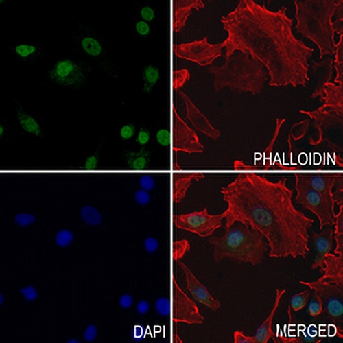
Immunofluorescent analysis of Histone H3 (Butyryl-K23) staining in A549 cells. Formalin-fixed cells were permeabilized with 0.1% Triton X-100 in TBS for 5-10 minutes and blocked with 3% BSA-PBS for 30 minutes at room temperature. Cells were probed with the primary antibody in 3% BSA-PBS and incubated overnight at 4 °C in a hidified chamber. Cells were washed with PBST and incubated with a AREX® Fluor 488 -conjugated secondary antibody (green) in PBS at room temperature in the dark. Phalloidin - AREX® Fluor 594 was used to stain Actin filaments (red). DAPI was used to stain the cell nuclei (blue).
FAQs
 New Products


