


HER3 Rabbit Monoclonal Antibody(ARB585)
 Datasheet
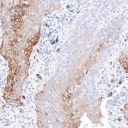
Key features and details
- Target:
- Clone ID:
- Source/Host:
- Reactivity:
- Applications:
- Dilution:
- Clonality:
- Storage:
-
Brand:

CAT.NO. : ARB6877
US$ Please choose
US$ Please choose
Size:
Trail, Bulk size or Custom requests Please contact us
 Product Details
Background
HER3 (Human Epidermal Growth Factor Receptor 3), a member of the EGFR tyrosine kinase receptor family, functions as a critical dimerization partner for other ERBB receptors (notably HER2) despite lacking intrinsic kinase activity. HER3 is predominantly expressed in kidney and tonsil. In tumor tissue, it is mainly expressed in breast cancer and lung cancer. HER3 is a critical biomarker and therapeutic target in oncology, with its overexpression and activation detected via immunohistochemistry (IHC), fluorescence in situ hybridization (FISH), or serum assays correlating strongly with aggressive tumor behavior, therapy resistance and poor prognosis. Pathologically, HER3+ tumor cells exhibit increased epithelialmesenchymal transition (EMT) signatures and stromal invasion.
Application
To ensure optimal assay performance, AREX recommends conducting reagent titration tailored to each testing system for optimal detection results.
*Results are sample-specific. Please refer to your local assay conditions and test parameters for reference.
Application | Dilution Ratio |
IHC | 1:100 - 1:200 |
Overview
Predicted Molecular Wt | 148kDa |
Purity | ProA affinity purified IgG |
Subcellular location | Membrane and cytoplasm |
Swissprot ID | P21860 |
Immunogen | Synthetic peptide. |
Storage Buffer | PBS 59%, Sodium azide 0.01%, Glycerol 40%, BSA 0.05% |
Recommended Method | Heat induced epitope retrieval with Tris-EDTA buffer (pH 9.0), primary antibody incubate at RT (18℃-25℃) for 30 minutes. |
Alternative Names | ErbB3, Receptor tyrosine-protein kinase erbB-3 |
Gene Symbol | ERBB3 |
Entrez Gene | 2065 |
*Clone Number, Reactivity, Source/Host and Clonality can be found in the product name and Key Features section above.
Data
Immunohistochemical staining of human lung tissue using HER3 Rabbit Monoclonal Antibody(ARB585).
Storage
Store at 4°C short term. For long term storage, store at -20°C, avoiding freeze/thaw cycles.
Note
For Research Use Only. Not for diagnostic, therapeutics, prophylactic or in vivo use.
FAQs
Are the pathology antibodies provided by AREX raw antibodies or ready-to-use solutions?

AREX Biosciences specializes in supplying high-quality IHC pathology raw antibodies (Raw Antibodies). We do not manufacture ready-to-use working solutions or IVD diagnostic reagents. We mainly provide concentrated raw materials to pathology reagent manufacturers and diagnostic platform companies to support product development and OEM production.
What development scenarios are pathology raw antibodies mainly used for?
Our raw antibodies are primarily used for the research, performance optimization, validation, and commercial-scale production of pathology IHC detection reagents. They are also suitable for companion diagnostics (CDx) projects and antibody screening.
How can I evaluate whether a raw antibody is suitable for our staining platform?
It is recommended to focus on specificity, sensitivity, background control, and performance on your target automated platforms (such as Roche Ventana, Leica Bond, Agilent Dako, etc.). We can provide internal test data for reference, but we recommend partners perform actual validation on their own platforms.
Can you provide samples for platform validation?
Yes. We can provide validation samples depending on the specific product. Some products may be provided free of charge, while others may involve a small sample fee. Handling fee and shipping fee will be charged separately. Please contact us for detailed confirmation.
How should raw antibodies be paired with detection systems?
AREX raw antibodies can be used with various polymer detection systems and ancillary reagents. We recommend optimization in combination with the ARExVisual® system or your existing detection platform to achieve better signal intensity and background control.
New Products


