


HER2 Mouse Monoclonal Antibody(C2141)
 Datasheet
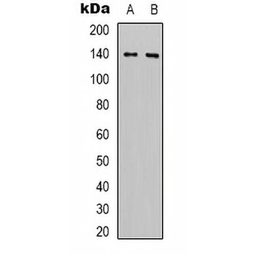
Key features and details
- Target:
- Source/Host:
- Reactivity:
- Clonality:
- Applications:
- Conjugation:
- Storage:
-
Brand:

 Product Details
WB | 1:2000 - 1:5000 |
IHC | 1:100 - 1:200 |
Description | Mouse monoclonal antibody to HER2 |
Specificity | Recognizes endogenous levels of HER2 protein. |
Antibody Type | Primary antibody |
Imnunogen | KLH-conjugated synthetic peptide encompassing a sequence of human HER2. The exact sequence is proprietary. |
Purification | Affinity chromatography |
Molecular Weight | Predicted: 137 kD; Observed: 185 kD |
Form/Buffer | Liquid in 0.42% Potassium phosphate, 0.87% Sodium chloride, pH 7.3, 30% glycerol, and 0.01% sodium azide. |
Alternative Names | HER2; MLN19; NEU; NGL; Receptor tyrosine-protein kinase erbB-2; Metastatic lymph node gene 19 protein; MLN 19; Proto-oncogene Neu; Proto-oncogene c-ErbB-2; Tyrosine kinase-type cell surface receptor HER2; p185erbB2; CD340 |
Gene Symbol | ERBB2 |
Entrez Gene | 2064(Human); 13866(Mouse) |
SwissProt | P04626(Human); P70424(Mouse); P06494(Rat) |
*Clone Number, Reactivity, Source/Host and Clonality can be found in the product name and Key Features section above.
Western blot analysis of HER2 expression in Hela (A), mouse brain (B) whole cell lysates. (Predicted band size: 137 kD; Observed band size: 185 kD)
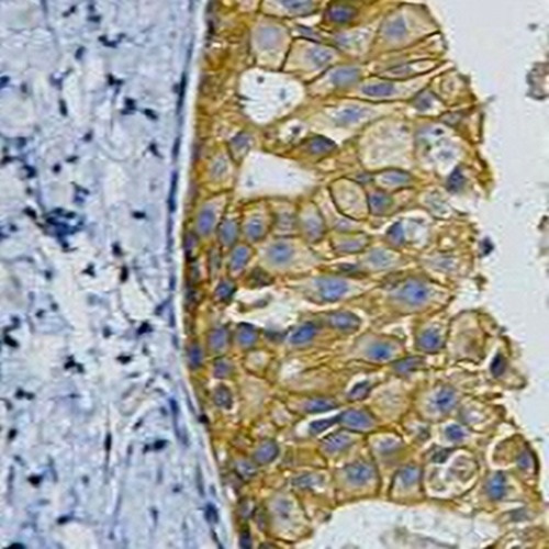
Immunohistochemical analysis of HER2 staining in human breast cancer formalin fixed paraffin embedded tissue section. The section was pre-treated using heat mediated antigen retrieval with sodium citrate buffer (pH 6.0). The section was then incubated with the antibody at room temperature and detected using an HRP conjugated compact polymer system. DAB was used as the chromogen. The section was then counterstained with haematoxylin and mounted with DPX.
FAQs
 New Products


