


H3K27M Rabbit Monoclonal Antibody(ARB964)
 Datasheet
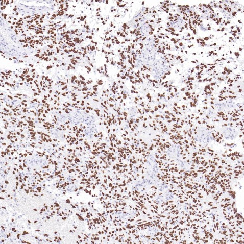
Key features and details
- Target:
- Clone ID:
- Source/Host:
- Reactivity:
- Applications:
- Dilution:
- Clonality:
- Storage:
-
Brand:

CAT.NO. : ARB6756
US$ Please choose
US$ Please choose
Size:
Trail, Bulk size or Custom requests Please contact us
 Product Details
Background
H3K27M: encoded by the mutation of a histone H3 gene such as H3F3A. It expressed during S phase, then expression strongly decreases as cell division slows down during the process of differentiation. Histones play a central role in transcription regulation, DNA repair, DNA replication and chromosomal stability.Diffuse midline glioma, H3K27M - mutant is a lethal brain tumor located in the thalamus, brain stem, or spinal cord. H3K27M plays a pivotal role in the tumorigenesis of this type of glioma. Some results indicated that the H3F3A K27M mutation was found in approximately 80% of diffuse intrinsic pontine gliomas (DIPGs), 50% of thalamic tumors, and 60% of spinal tumors.H3K27M can be used as an important molecular marker in the diagnosis of diffuse midline gliomas in children and young adults, it is associated with poor prognosis.
Application
To ensure optimal assay performance, AREX recommends conducting reagent titration tailored to each testing system for optimal detection results.
*Results are sample-specific. Please refer to your local assay conditions and test parameters for reference.
Application | Dilution Ratio |
IHC | 1:100 - 1:200 |
Overview
Predicted Molecular Wt | 15kDa |
Purity | ProA affinity purified IgG |
Subcellular location | Nucleus |
Swissprot ID | P68431 |
Immunogen | Synthetic peptide. |
Storage Buffer | PBS 59%, Sodium azide 0.01%, Glycerol 40%, BSA 0.05% |
Recommended Method | Heat induced epitope retrieval with Tris-EDTA buffer (pH 9.0), primary antibody incubate at RT (18℃-25℃) for 30 minutes |
Alternative Names | PHH3, Mitosis marker |
Gene Symbol | H3C1 (HIST1H3A) |
Entrez Gene | 8350 |
*Clone Number, Reactivity, Source/Host and Clonality can be found in the product name and Key Features section above.
Data
Immunohistochemical staining of human diffuse midline glioma tissue using H3K27M Rabbit Monoclonal Antibody(ARB964).
Storage
Store at 4°C short term. For long term storage, store at -20°C, avoiding freeze/thaw cycles.
Note
For Research Use Only. Not for diagnostic, therapeutics, prophylactic or in vivo use.
FAQs
Are the pathology antibodies provided by AREX raw antibodies or ready-to-use solutions?

AREX Biosciences specializes in supplying high-quality IHC pathology raw antibodies (Raw Antibodies). We do not manufacture ready-to-use working solutions or IVD diagnostic reagents. We mainly provide concentrated raw materials to pathology reagent manufacturers and diagnostic platform companies to support product development and OEM production.
What development scenarios are pathology raw antibodies mainly used for?
Our raw antibodies are primarily used for the research, performance optimization, validation, and commercial-scale production of pathology IHC detection reagents. They are also suitable for companion diagnostics (CDx) projects and antibody screening.
How can I evaluate whether a raw antibody is suitable for our staining platform?
It is recommended to focus on specificity, sensitivity, background control, and performance on your target automated platforms (such as Roche Ventana, Leica Bond, Agilent Dako, etc.). We can provide internal test data for reference, but we recommend partners perform actual validation on their own platforms.
Can you provide samples for platform validation?
Yes. We can provide validation samples depending on the specific product. Some products may be provided free of charge, while others may involve a small sample fee. Handling fee and shipping fee will be charged separately. Please contact us for detailed confirmation.
How should raw antibodies be paired with detection systems?
AREX raw antibodies can be used with various polymer detection systems and ancillary reagents. We recommend optimization in combination with the ARExVisual® system or your existing detection platform to achieve better signal intensity and background control.
New Products


