


GTF2I Rabbit Polyclonal Antibody
 Datasheet
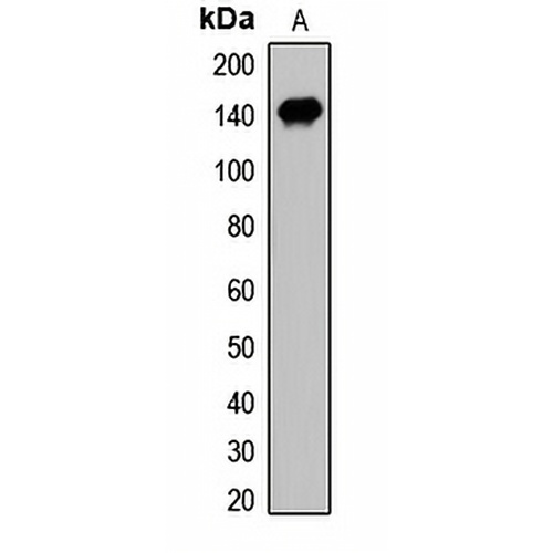
Key features and details
- Target:
- Source/Host:
- Reactivity:
- Clonality:
- Applications:
- Conjugation:
- Storage:
-
Brand:

 Product Details
WB | 1:500 - 1:2000 |
IF/ICC | 1:50 - 1:200 |
Description | Rabbit polyclonal antibody to GTF2I |
Specificity | Recognizes endogenous levels of GTF2I protein. |
Antibody Type | Primary antibody |
Imnunogen | KLH-conjugated synthetic peptide of human GTF2I |
Purification | The antibody was purified by immunogen affinity chromatography. |
Molecular Weight | Predicted: 30; Observed: 135 kD |
Form/Buffer | Liquid in 0.42% Potassium phosphate, 0.87% Sodium chloride, pH 7.3, 30% glycerol, and 0.01% sodium azide. |
Alternative Names | BAP135; WBSCR6; General transcription factor II-I; GTFII-I; TFII-I; Bruton tyrosine kinase-associated protein 135; BAP-135; BTK-associated protein 135; SRF-Phox1-interacting protein; SPIN; Williams-Beuren syndrome chromosomal region 6 protein |
Gene Symbol | GTF2I |
Entrez Gene | 2969(Human); 14886(Mouse); 353256(Rat) |
SwissProt | P78347(Human); Q9ESZ8(Mouse); Q5U2Y1(Rat) |
*Clone Number, Reactivity, Source/Host and Clonality can be found in the product name and Key Features section above.
Western blot analysis of GTF2I expression in HEK293T (A) whole cell lysates. (Predicted band size: 30; 107; 110; 112 kD; Observed band size: 135 kD)
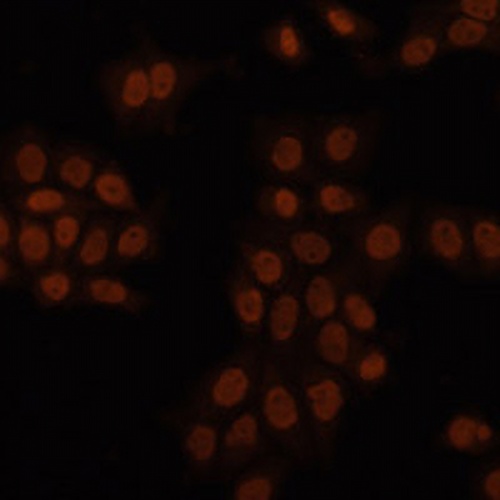
Immunofluorescent analysis of GTF2I staining in HeLa cells. Formalin-fixed cells were permeabilized with 0.1% Triton X-100 in TBS for 5-10 minutes and blocked with 3% BSA-PBS for 30 minutes at room temperature. Cells were probed with the primary antibody in 3% BSA-PBS and incubated overnight at 4 °C in a humidified chamber. Cells were washed with PBST and incubated with a AREX® Fluor 594-conjugated secondary antibody (red) in PBS at room temperature in the dark. DAPI was used to stain the cell nuclei (blue).
FAQs
 New Products


