


GEF H1 Rabbit Polyclonal Antibody
 Datasheet
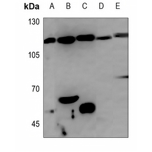
Key features and details
- Target:
- Source/Host:
- Reactivity:
- Clonality:
- Applications:
- Conjugation:
- Storage:
-
Brand:

 Product Details
WB | 1:500 - 1:1000 |
IHC | 1:50 - 1:100 |
Description | Rabbit polyclonal antibody to GEF H1 |
Specificity | Recognizes endogenous levels of GEF H1 protein. |
Antibody Type | Primary antibody |
Imnunogen | KLH-conjugated synthetic peptide encompassing a sequence within the center region of human GEF H1. The exact sequence is proprietary. |
Purification | The antibody was purified by immunogen affinity chromatography. |
Molecular Weight | Predicted: 111 kD; Observed: 111 kD |
Form/Buffer | Liquid in 0.42% Potassium phosphate, 0.87% Sodium chloride, pH 7.3, 30% glycerol, and 0.01% sodium azide. |
Alternative Names | KIAA0651; LFP40; Rho guanine nucleotide exchange factor 2; Guanine nucleotide exchange factor H1; GEF-H1; Microtubule-regulated Rho-GEF; Proliferating cell nucleolar antigen p40 |
Gene Symbol | ARHGEF2 |
Entrez Gene | 9181(Human); 16800(Mouse); 310635(Rat) |
SwissProt | Q92974(Human); Q60875(Mouse); Q5FVC2(Rat) |
*Clone Number, Reactivity, Source/Host and Clonality can be found in the product name and Key Features section above.
Western blot analysis of GEF H1 expression in SHSY5Y (A), mouse liver (B), mouse testis (C), rat spleen (D), rat testis (E) whole cell lysates. (Predicted band size: 111 kD; Observed band size: 111 kD)
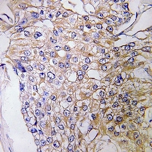
Immunohistochemical analysis of GEF H1 staining in human breast cancer formalin fixed paraffin embedded tissue section. The section was pre-treated using heat mediated antigen retrieval with sodium citrate buffer (pH 6.0). The section was then incubated with the antibody at room temperature and detected using an HRP conjugated compact polymer system. DAB was used as the chromogen. The section was then counterstained with haematoxylin and mounted with DPX.
FAQs
 New Products


