


CPT1A Rabbit Monoclonal Antibody(C4067)


Key features and details
- Reactivity:
- Application:
- Host:
- Clonality:
- Target:
-
Brand:

CAT.NO. : AMA03887
US$ Please choose
US$ Please choose
 Product Details
Background
The mitochondrial oxidation of long-chain fatty acids is initiated by the sequential action of carnitine palmitoyltransferase I (which is located in the outer membrane and is detergent-labile) and carnitine palmitoyltransferase II (which is located in the inner membrane and is detergent-stable), together with a carnitine-acylcarnitine translocase. CPT I is the key enzyme in the carnitine-dependent transport across the mitochondrial inner membrane and its deficiency results in a decreased rate of fatty acid beta-oxidation. Alternatively spliced transcript variants encoding different isoforms have been found for this gene.
Application
|
Application |
Dilution Ratio |
|
WB |
1:500-1:2000 |
|
IF/ICC |
1:50-1:200 |
Overview
|
Product Description |
Recombinant rabbit monoclonal antibody to CPT1A |
|
Immunogen |
KLH-conjugated synthetic peptide encompassing a sequence within human CPT1A protein. The exact sequence is proprietary. |
|
Purification Method |
The antibody was purified by immunogen affinity chromatography. |
|
Clonality |
Monoclonal |
|
Form |
Liquid in PBS, pH 7.3, 50% glycerol, 0.05% BSA, and 0.05% Proclin300. |
|
Gene Symbol |
CPT1A |
|
Alternative Names |
CPT1; Carnitine O-palmitoyltransferase 1, liver isoform; CPT1-L; Carnitine O-palmitoyltransferase I, liver isoform; CPT I; CPTI-L; Carnitine palmitoyltransferase 1A |
|
Gene ID (Human) |
1374 |
|
Gene ID (Mouse) |
12894 |
|
Gene ID (Rat) |
25757 |
|
Protein ID (Human) |
P50416 |
|
Protein ID (Mouse) |
P97742 |
|
Protein ID (Rat) |
P32198 |
Data

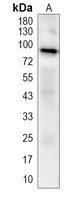
Western blot analysis of CPT1A expression in HeLa (A) whole cell lysates. (Predicted band size: 86; 88 kD; Observed band size: 88 kD)
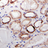
Immunohistochemical analysis of CPT1A staining in human kidney formalin fixed paraffin embedded tissue section. The section was pre-treated using heat mediated antigen retrieval with sodium citrate buffer (pH 6.0). The section was then incubated with the antibody at room temperature and detected using an HRP conjugated compact polymer system. DAB was used as the chromogen. The section was then counterstained with haematoxylin and mounted with DPX.
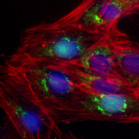
Immunofluorescent analysis of CPT1A staining in SKOV3 cells. Formalin-fixed cells were permeabilized with 0.1% Triton X-100 in TBS for 5-10 minutes and blocked with 3% BSA-PBS for 30 minutes at room temperature. Cells were probed with the primary antibody in 3% BSA-PBS and incubated overnight at 4 °C in a humidified chamber. Cells were washed with PBST and incubated with a AREX®Flour 594-conjugated secondary antibody (red) in PBS at room temperature in the dark. DAPI was used to stain the cell nuclei (blue).
Storage
Store at 4°C short term. For long term storage, store at -20°C, avoiding freeze/thaw cycles.
Research Use Only
For Research Use Only. Not for use in diagnostic procedures.
New Products


