


CD79a Rabbit Monoclonal Antibody(ARA771)
 Datasheet
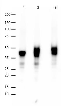
Key features and details
- Target:
- Source/Host:
- Reactivity:
- Clonality:
- Applications:
- Conjugation:
- Storage:
-
Brand:

 Product Details
WB | 1:1000-1:2000 |
IHC | 1:6400-1:12800 |
FC | 1:800-1:2000 |
Description | Rabbit Monoclonal Antibody to CD79a |
Antibody Type | Primary antibody |
Predicted MW | 25kDa |
Immunogen | A synthetic peptide corresponding to the N-term of CD79a was used as an immunogen. |
Purification | ProA affinity purified IgG |
Form/Buffer | PBS 59%, Sodium azide 0.01%, Glycerol 40%, BSA 0.46%. |
Alternative Names | IGA; MB1; B-cell antigen receptor complex-associated protein alpha chain; Ig-alpha; MB-1 membrane glycoprotein; Membrane-bound immunoglobulin-associated protein; Surface IgM-associated protein; CD79a |
Gene Symbol | CD79A |
Entrez Gene | 973(Human) |
Swissprot | P11912 |
*Clone Number, Reactivity, Source/Host and Clonality can be found in the product name and Key Features section above.
All lanes: Anti-CD79a antibody at 1:1,000 dilution
Predicted MW: 25 kDa
Observed MW: 45-48 kDa
Lane 1: Raji
Lane 2: Ramos
Lane 3: Daudi
Lysate at 10 µg per lane
2nd Ab:
GAR HRP(H+L) 1:5,000
Exposure: 20s
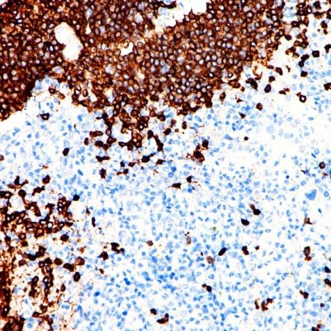
Immunohistochemistry (Formalin/PFA-fixed paraffin-embedded sections) analysis of tonsil tissue labelling CD79a with ARA771 at 1:12,800. Heat mediated antigen retrieval was performed using Tris/EDTA buffer pH 9.0.
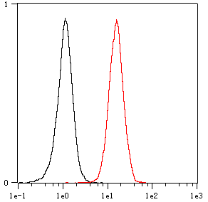
Overlay histogram showing Ramos cells stained with ARA771 (Red). The cells were fixed with 4% paraformaldehyde (10 min) and then permeabilized with 0.1% TritonX-100 for 15 min. The cells were then incubated in the antibody (ARA771, 1:2,000 dilution) in 1x PBS/1% BSA for 30 min at room temperature. The secondary antibody used was a Goat Anti-Rabbit Alexa Fluor<sup>®</sup> 488 (IgG H+L) at 1:2,000 dilution for 20 min at room temperature. Unlabelled sample (Black) was used as a control.
FAQs
 New Products


