


CD4 Rabbit Monoclonal Antibody(ARA818)
 Datasheet
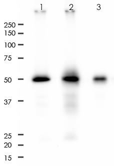
Key features and details
- Target:
- Source/Host:
- Reactivity:
- Clonality:
- Applications:
- Conjugation:
- Storage:
-
Brand:

 Product Details
WB | 1:2000-1:5000 |
IHC | 1:1000-1:2000 |
FC | 1:10-1:50 |
IP | 1:50 |
Description | Rabbit Monoclonal Antibody to CD4 |
Antibody Type | Primary antibody |
Predicted MW | 51kDa |
Immunogen | Recombinant protein corresponding to aa200-400 of human CD4 was used as an immunogen. |
Purification | ProA affinity purified IgG |
Form/Buffer | PBS 59%, Sodium azide 0.01%, Glycerol 40%, BSA 0.93%. |
Alternative Names | T-cell surface glycoprotein CD4; T-cell surface antigen T4/Leu-3; CD4 |
Gene Symbol | CD4 |
Entrez Gene | 920(Human) |
Swissprot | P01730 |
*Clone Number, Reactivity, Source/Host and Clonality can be found in the product name and Key Features section above.
All lanes: Anti-CD4 antibody at 1:2,000 dilution
Predicted MW: 51 kDa
Observed MW: 51 kDa
Lane 1: Molt-4
Lane 2: THP-1
Lane 3: HuT-78
Lysate at 10 µg per lane
2nd Ab:
G&R HRP(H+L) 1:10,000
Exposure: 100s
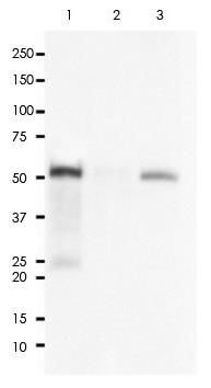
CD4 was immunoprecipitated from 0.4mg of Molt-4 whole cell lysate with ARA818 at 1:50 dilution.
2nd Ab:
GAR HRP for IP 1:500
Lane 1: ARA818 IP in Molt-4 whole cell lysate
Lane 2: Rabbit IgG instead of RR648 in Molt-4 whole cell lysate
Lane 3: Molt-4 whole cell lysate, 10 µg (input)
Exposure: 120s
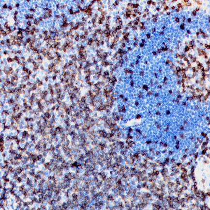
Immunohistochemistry (Formalin/PFA-fixed paraffin-embedded sections) analysis of human colon tissue labelling CD4 with ARA818 at 1:2,000. Heat mediated antigen retrieval was performed using Tris/EDTA buffer pH 9.0.
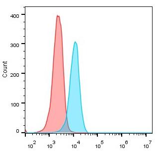
Overlay histogram showing Jurkat cells stained with ARA818 (Blue). The cells were fixed with 4% paraformaldehyde for 10 min. The cells were then incubated in the antibody (ARA818, 1:50 dilution) in 1x PBS/1% BSA for 30 min at room temperature. The secondary antibody used was a Goat Anti-Rabbit Alexa Fluor<sup>®</sup> 488 (IgG H+L) at 1:2,000 dilution for 20 min at room temperature. Unlabelled sample (Red) was used as a control.
FAQs
 New Products


