


CD1a Rabbit Monoclonal Antibody(ARB894)
 Datasheet
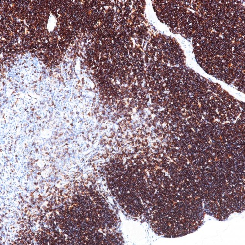
Key features and details
- Target:
- Clone ID:
- Source/Host:
- Reactivity:
- Applications:
- Dilution:
- Clonality:
- Storage:
-
Brand:

CAT.NO. : ARB6686
US$ Please choose
US$ Please choose
Size:
Trail, Bulk size or Custom requests Please contact us
 Product Details
Background
CD1a is a member of the family group 1 CD1 proteins (CD1a, CD1b, and CD1c), which share the capacity to present microbial lipid antigens to cells.CD1a is expressed predominantly on Langerhans cells (LCs), it can be expressed by T-cells under certain conditions. Immature thymocytes express CD1a, as do some T-cells involved in neoplastic conditions such as pre-lymphoblastic lymphomas/leukemias and follicular dendritic cell sarcoma.CD1a expression on pathogenic Langerhans cells (LCs) in LCH is regarded as a hallmark of this disease and may as a potential target for future therapeutic strategies against inflammatory skin diseases.
Application
To ensure optimal assay performance, AREX recommends conducting reagent titration tailored to each testing system for optimal detection results.
*Results are sample-specific. Please refer to your local assay conditions and test parameters for reference.
Application | Dilution Ratio |
IHC | 1:100 - 1:200 |
Overview
Predicted Molecular Wt | 37 kDa |
Purity | ProA affinity purified IgG |
Subcellular location | Membrane |
Swissprot ID | P06126 |
Immunogen | Synthetic peptide corresponding to CD1a residues within aa1-100 |
Storage Buffer | PBS 59%, Sodium azide 0.01%, Glycerol 40%, BSA 0.05% |
Recommended method | Heat induced epitope retrieval with Tris-EDTA buffer (pH 9.0), primary antibody incubate at RT (18° C-25° C) for 30 minutes. |
Alternative Names | T-cell surface glycoprotein CD1a; T-cell surface antigen T6/Leu-6; hTa1 thymocyte antigen; CD1a |
Gene Symbol | CD1A |
Entrez Gene | 909 |
*Clone Number, Reactivity, Source/Host and Clonality can be found in the product name and Key Features section above.
Data
Immunohistochemical staining of human thymus tissue sections using CD1a Rabbit Monoclonal Antibody (ARB894).
Storage
Store at 4°C short term. For long term storage, store at -20°C, avoiding freeze/thaw cycles.
Note
For Research Use Only. Not for diagnostic, therapeutics, prophylactic or in vivo use.
FAQs
Are the pathology antibodies provided by AREX raw antibodies or ready-to-use solutions?

AREX Biosciences specializes in supplying high-quality IHC pathology raw antibodies (Raw Antibodies). We do not manufacture ready-to-use working solutions or IVD diagnostic reagents. We mainly provide concentrated raw materials to pathology reagent manufacturers and diagnostic platform companies to support product development and OEM production.
What development scenarios are pathology raw antibodies mainly used for?
Our raw antibodies are primarily used for the research, performance optimization, validation, and commercial-scale production of pathology IHC detection reagents. They are also suitable for companion diagnostics (CDx) projects and antibody screening.
How can I evaluate whether a raw antibody is suitable for our staining platform?
It is recommended to focus on specificity, sensitivity, background control, and performance on your target automated platforms (such as Roche Ventana, Leica Bond, Agilent Dako, etc.). We can provide internal test data for reference, but we recommend partners perform actual validation on their own platforms.
Can you provide samples for platform validation?
Yes. We can provide validation samples depending on the specific product. Some products may be provided free of charge, while others may involve a small sample fee. Handling fee and shipping fee will be charged separately. Please contact us for detailed confirmation.
How should raw antibodies be paired with detection systems?
AREX raw antibodies can be used with various polymer detection systems and ancillary reagents. We recommend optimization in combination with the ARExVisual® system or your existing detection platform to achieve better signal intensity and background control.
New Products


