


c-FER Mouse Monoclonal Antibody(C3856)
 Datasheet
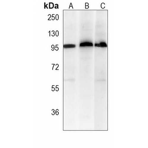
Key features and details
- Target:
- Source/Host:
- Reactivity:
- Clonality:
- Applications:
- Conjugation:
- Storage:
-
Brand:

 Product Details
WB | 1:500 - 1:2000 |
IF/ICC | 1:10 - 1:50 |
FC | 1:10 - 1:50 |
Description | Mouse monoclonal antibody to c-FER |
Specificity | Recognizes endogenous levels of c-FER protein. |
Antibody Type | Primary antibody |
Imnunogen | Recombinant fusion protein of human c-FER. The exact sequence is proprietary. |
Purification | This antibody is purified through a protein G column. |
Molecular Weight | Predicted: 94 kD; Observed: 100 kD |
Form/Buffer | Mouse IgG2a kappa. Liquid in PBS, pH 7.3, 30% glycerol, and 0.01% sodium azide. |
Alternative Names | TYK3; Tyrosine-protein kinase Fer; Feline encephalitis virus-related kinase FER; Fujinami poultry sarcoma/Feline sarcoma-related protein Fer; Proto-oncogene c-Fer; Tyrosine kinase 3; p94-Fer |
Gene Symbol | FER |
Entrez Gene | 14158(Mouse) |
SwissProt | P70451(Mouse) |
*Clone Number, Reactivity, Source/Host and Clonality can be found in the product name and Key Features section above.
Western blot analysis of c-FER expression in NIH3T3 (A), mouse testis (B), mouse liver (C) whole cell lysates. (Predicted band size: 94 kD; Observed band size: 100 kD)
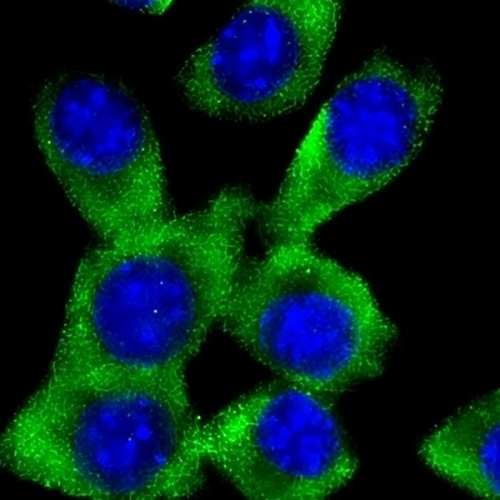
Immunofluorescent analysis of c-FER staining in NIH3T3 cells. Formalin-fixed cells were permeabilized with 0.1% Triton X-100 in TBS for 5-10 minutes and blocked with 3% BSA-PBS for 30 minutes at room temperature. Cells were probed with the primary antibody in 3% BSA-PBS and incubated overnight at 4 °C in a humidified chamber. Cells were washed with PBST and incubated with a AREX® Fluor 488 -conjugated secondary antibody (green) in PBS at room temperature in the dark. DAPI was used to stain the cell nuclei (blue).
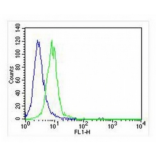
Flow cytometric analysis of NIH3T3 cells using Anti-c-FER Antibody. The cells were fixed with 2% paraformaldehyde (10 min) and then permeabilized with 90% methanol for 10 min. The cells were incubated in 2% bovine serum albumin to block non-specific protein-protein interactions followed by the antibody at 37 °C for 60 min. The secondary antibody Goat Anti-Mouse IgG (H&L) - AREX® Fluor 488 was incubated at 37 °C for 40 min. Isotype control antibody (blue line) was used under the same condition.
FAQs
 New Products


