


Aurora A Mouse Monoclonal Antibody(C3315)
 Datasheet
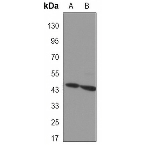
Key features and details
- Target:
- Source/Host:
- Reactivity:
- Clonality:
- Applications:
- Conjugation:
- Storage:
-
Brand:

 Product Details
WB | 1:500 - 1:1000 |
IF/ICC | 1:50 - 1:100 |
Description | Mouse monoclonal antibody to Aurora A |
Specificity | Recognizes endogenous levels of Aurora A protein. |
Antibody Type | Primary antibody |
Imnunogen | Purified recombinant human Aurora Kinase A protein fragments expressed in E.coli. |
Purification | The antibody was purified by immunogen affinity chromatography. |
Molecular Weight | Predicted: 46 kD; Observed: 46 kD |
Form/Buffer | Liquid in PBS containing 50% glycerol, 0.5% BSA and 0.02% sodium azide, pH 7.3. |
Alternative Names | AIK; AIRK1; ARK1; AURA; AYK1; BTAK; IAK1; STK15; STK6; Aurora kinase A; Aurora 2; Aurora/IPL1-related kinase 1; ARK-1; Aurora-related kinase 1; hARK1; Breast tumor-amplified kinase; Serine/threonine-protein kinase 15; Serine/threonine-protein kinase 6; Serine/threonine-protein kinase aurora-A |
Gene Symbol | AURKA |
Entrez Gene | 6790(Human) |
SwissProt | O14965(Human) |
*Clone Number, Reactivity, Source/Host and Clonality can be found in the product name and Key Features section above.
Western blot analysis of Aurora A expression in SW480 (A), COS7 (B) whole cell lysates. (Predicted band size: 46 kD; Observed band size: 46 kD)
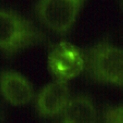
Immunofluorescent analysis of Aurora A staining in HeLa cells. Formalin-fixed cells were permeabilized with 0.1% Triton X-100 in TBS for 5-10 minutes and blocked with 3% BSA-PBS for 30 minutes at room temperature. Cells were probed with the primary antibody in 3% BSA-PBS and incubated overnight at 4 °C in a hidified chamber. Cells were washed with PBST and incubated with a AREX® Fluor 488 -conjugated secondary antibody (green) in PBS at room temperature in the dark.
FAQs
 New Products


