Focal Adhesion Kinase Mouse Monoclonal Antibody(C3366)
 Datasheet
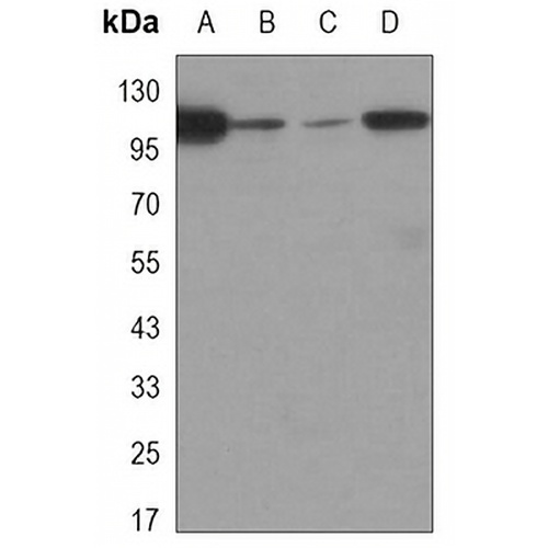
Key features and details
- Target:
- Source/Host:
- Reactivity:
- Clonality:
- Applications:
- Conjugation:
- Storage:
-
Brand:

 Product Details
WB | 1:500 - 1:1000 |
IF/ICC | 1:50 - 1:100 |
IP | 1:10 - 1:50 |
Description | Mouse monoclonal antibody to Focal Adhesion Kinase |
Specificity | Recognizes endogenous levels of Focal Adhesion Kinase protein. |
Antibody Type | Primary antibody |
Imnunogen | Purified recombinant human FAK protein fragments expressed in E.coli. |
Purification | The antibody was purified by immunogen affinity chromatography. |
Molecular Weight | Predicted: 119; Observed: 125 kD |
Form/Buffer | Liquid in PBS containing 50% glycerol, 0.5% BSA and 0.02% sodium azide, pH 7.3. |
Alternative Names | FAK; FAK1; Focal adhesion kinase 1; FADK 1; Focal adhesion kinase-related nonkinase; FRNK; Protein phosphatase 1 regulatory subunit 71; PPP1R71; Protein-tyrosine kinase 2; p125FAK; pp125FAK |
Gene Symbol | PTK2 |
Entrez Gene | 5747(Human) |
SwissProt | Q05397(Human) |
*Clone Number, Reactivity, Source/Host and Clonality can be found in the product name and Key Features section above.
Western blot analysis of Focal Adhesion Kinase expression in COS7 (A), HUVEC (B), A549 (C), Hela (D) whole cell lysates. (Predicted band size: 119; 99; 63; 48; 120; 39; 114 kD; Observed band size: 125 kD)
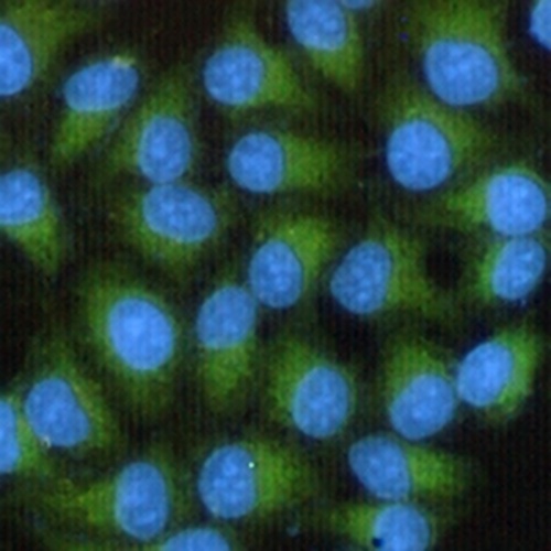
Immunofluorescent analysis of Focal Adhesion Kinase staining in HeLa cells. Formalin-fixed cells were permeabilized with 0.1% Triton X-100 in TBS for 5-10 minutes and blocked with 3% BSA-PBS for 30 minutes at room temperature. Cells were probed with the primary antibody in 3% BSA-PBS and incubated overnight at 4 °C in a hidified chamber. Cells were washed with PBST and incubated with a AREX® Fluor 488 -conjugated secondary antibody (green) in PBS at room temperature in the dark. DAPI was used to stain the cell nuclei (blue).
FAQs
 New Products