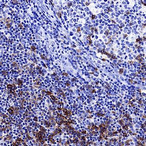
Caspase-3 (pro+p17) Rabbit Monoclonal Antibody(ARA793)
 Datasheet
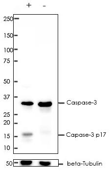
Key features and details
- Target:
- Source/Host:
- Reactivity:
- Clonality:
- Applications:
- Conjugation:
- Storage:
-
Brand:

 Product Details
WB | 1:1000-1:2000 |
IF/ICC | 1:200-1:800 |
FC | 1:200-1:1000 |
IP | 1:50 |
Description | Rabbit Monoclonal Antibody to Caspase-3 (pro+p17) |
Antibody Type | Primary antibody |
Predicted MW | 32/17kDa |
Immunogen | A synthetic peptide corresponding to aa1-100 of human Caspase-3 was used as an immunogen. |
Purification | ProA affinity purified IgG |
Form/Buffer | PBS 59%, Sodium azide 0.01%, Glycerol 40%, BSA 0.68%. |
Alternative Names | CPP32; Caspase-3; CASP-3; Apopain; Cysteine protease CPP32; CPP-32; Protein Yama; SREBP cleavage activity 1; SCA-1 |
Gene Symbol | CASP3 |
Entrez Gene | 836(Human); 12367(Mouse); 25402(Rat) |
Swissprot | P42574 |
*Clone Number, Reactivity, Source/Host and Clonality can be found in the product name and Key Features section above.
All lanes: Anti-Caspase-3 antibody at 1:2,000 dilution
Predicted MW: 32/17 kDa
Observed MW: 35/17 kDa
Lane +: Jurkat treated with 1 uM staurosporine for 4h
Lane -: Jurkat untreated
Lysate at 20 µg per lane
2nd Ab:
GAR HRP(H+L) 1:5,000
Exposure: 20s
This antibody is specific for the pro form and the p17 cleaved form of
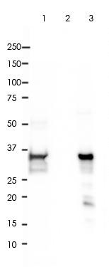
Caspase-3 was immunoprecipitated from 0.4mg of Jurkat whole cell lysate with ARA793 at 1:50 dilution.
2nd Ab:
GAR HRP for IP 1:500
Lane 1: ARA793 IP in Jurkat whole cell lysate
Lane 2: PBS instead of ARA793 in Jurkat whole cell lysate
Lane 3: Jurkat whole cell lysate, 10 µg (input)
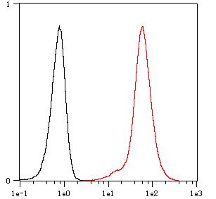
Overlay histogram showing Ramos cells stained with ARA793 (Red). The cells were fixed with 4% paraformaldehyde (10 min) and then permeabilized with 0.1% TritonX-100 for 15 min. The cells were then incubated in the antibody (ARA793, 1:1,000 dilution) in 1x PBS/1% BSA for 30 min at room temperature. The secondary antibody used was a Goat Anti-Rabbit Alexa Fluor<sup>®</sup> 488 (IgG H+L) at 1:2,000 dilution for 20 min at room temperature. Unlabelled sample (Black) was used as a control.
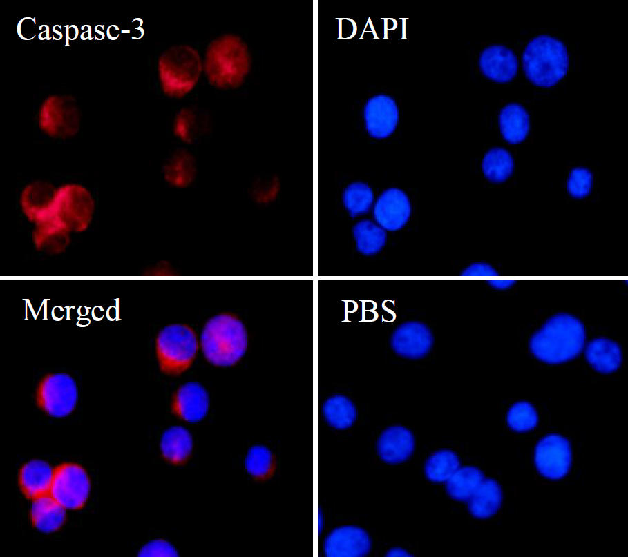
ARA793 staining Caspase-3 in Jurkat cells by IF/ICC (immunofluorescence/immunocytochemistry). Cells were fixed with paraformaldehyde, permeabilized with 0.1% Triton X-100 and blocked with 10% goat serum for half an hour at room temperature. Samples were incubated with primary antibody (1:800) at 4°C. An Alexa Fluor<sup>®</sup> 594-conjugated Goat Anti-Rabbit IgG polyclonal was used as the secondary antibody (1:500). DAPI (blue) was used as the nuclear counter stain.
Control: PBS and secondary antibody, An Alexa Fluor<sup>®</sup> 488-conjugated Goat Anti-Rabbit IgG (1:500).
FAQs
 New Products