c-Myc Rabbit Monoclonal Antibody(C3088)
 Datasheet
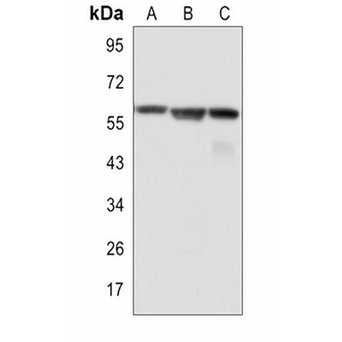
Key features and details
- Target:
- Source/Host:
- Reactivity:
- Clonality:
- Applications:
- Conjugation:
- Storage:
-
Brand:

 Product Details
WB | 1:500 - 1:1000 |
IF/ICC | 1:50 - 1:100 |
IP | 1:10 - 1:50 |
Description | Rabbit monoclonal antibody to c-Myc |
Specificity | Recognizes endogenous levels of c-Myc protein. |
Antibody Type | Primary antibody |
Imnunogen | A synthetic peptide of human c-Myc |
Purification | The antibody was purified by immunogen affinity chromatography. |
Molecular Weight | Predicted: 49 kD; Observed: 57-65 kD |
Form/Buffer | Liquid in 50mM Tris-Glycine (pH 7.4), 0.15M NaCl, 50% Glycerol, 0.01% Sodium azide and 0.05% BSA. |
Alternative Names | BHLHE39; Myc proto-oncogene protein; Class E basic helix-loop-helix protein 39; bHLHe39; Proto-oncogene c-Myc; Transcription factor p64 |
Gene Symbol | MYC |
Entrez Gene | 4609(Human); 17869(Mouse); 24577(Rat) |
SwissProt | P01106(Human); P01108(Mouse); P09416(Rat) |
*Clone Number, Reactivity, Source/Host and Clonality can be found in the product name and Key Features section above.
Western blot analysis of c-Myc expression in K562 (A), C6 (B), NIH3T3 (C) whole cell lysates. (Predicted band size: 49 kD; Observed band size: 57-65 kD)
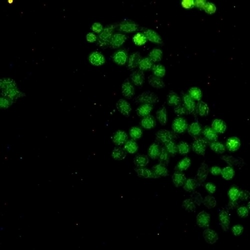
Immunofluorescent analysis of c-Myc staining in HeLa cells. Formalin-fixed cells were permeabilized with 0.1% Triton X-100 in TBS for 5-10 minutes and blocked with 3% BSA-PBS for 30 minutes at room temperature. Cells were probed with the primary antibody in 3% BSA-PBS and incubated overnight at 4 °C in a hidified chamber. Cells were washed with PBST and incubated with a AREX® Fluor 488 -conjugated secondary antibody (green) in PBS at room temperature in the dark.
FAQs
 New Products