AP2 alpha/beta Rabbit Polyclonal Antibody

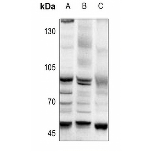
Key features and details

Rabbit polyclonal antibody to AP2 alpha/beta
  • Target: AP2 alpha/beta
  • Source/Host: Rabbit
  • Reactivity: Human, Mouse, Rat, Chicken, Pig, Sheep
  • Clonality: Polyclonal
  • Applications: WB, IHC, IF/ICC, ChIP, EMSA
  • Conjugation: Unconjugated
  • Storage: at-20°C
  • Brand:
CAT.NO. : APA07624
US$ Please choose
US$ Please choose
Size:
Trail, Bulk size or Custom requests Please contact us
Product Details
Background
Sequence-specific DNA-binding protein that interacts with inducible viral and cellular enhancer elements to regulate transcription of selected genes. AP-2 factors bind to the consensus sequence 5'-GCCNNNGGC-3' and activate genes involved in a large spectrum of important biological functions including proper eye, face, body wall, limb and neural tube development. They also suppress a number of genes including MCAM/MUC18, C/EBP alpha and MYC. AP-2-alpha is the only AP-2 protein required for early morphogenesis of the lens vesicle. Together with the CITED2 coactivator, stimulates the PITX2 P1 promoter transcription activation. Associates with chromatin to the PITX2 P1 promoter region.
Application
To ensure optimal assay performance, AREX recommends conducting reagent titration tailored to each testing system for optimal detection results.

WB

1:500 - 1:1000

IHC

1:100 - 1:200

IF/ICC

1:100 - 1:500

ChIP

1:100 - 1:500

EMSA

Use at an assay dependent dilution

*Results are sample-specific. Please refer to your local assay conditions and test parameters for reference.
Overview

Description

Rabbit polyclonal antibody to AP2 alpha/beta

Specificity

Recognizes endogenous levels of AP2 alpha/beta protein.

Antibody Type

Primary antibody

Imnunogen

KLH-conjugated synthetic peptide encompassing a sequence within the C-term region of human AP2 alpha/beta. The exact sequence is proprietary.

Purification

The antibody was purified by immunogen affinity chromatography.

Molecular Weight

Predicted: 48; Observed: 48 kD

Form/Buffer

Liquid in 0.42% Potassium phosphate, 0.87% Sodium chloride, pH 7.3, 30% glycerol, and 0.01% sodium azide.

Alternative Names

TFAP2A; AP2TF; TFAP2; Transcription factor AP-2-alpha; AP2-alpha; AP-2 transcription factor; Activating enhancer-binding protein 2-alpha; Activator protein 2; AP-2; TFAP2B; Transcription factor AP-2-beta; AP2-beta; Activating enhancer-binding protein 2-beta

Gene Symbol

TFAP2A; TFAP2B

Entrez Gene

7020(Human); 21418; 21419(Mouse)

SwissProt

P05549; Q92481(Human); P34056; Q61313(Mouse); P58197(Rat)

*AREX continuously optimizes our products. Webpage content may not reflect the latest updates. For inquiries, please contact info@arexbio.com or your local distributor.
*Clone Number, Reactivity, Source/Host and Clonality can be found in the product name and Key Features section above.
Data

Western blot analysis of AP2 alpha/beta expression in PC3 (A), MCF7 (B), Myla2059 (C) whole cell lysates. (Predicted band size: 48; 50 kD; Observed band size: 48 kD)

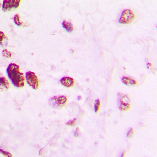
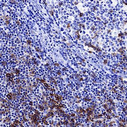
Immunohistochemical analysis of AP2 alpha/beta staining in human lung cancer formalin fixed paraffin embedded tissue section. The section was pre-treated using heat mediated antigen retrieval with sodium citrate buffer (pH 6.0). The section was then incubated with the antibody at room temperature and detected using an HRP conjugated compact polymer system. DAB was used as the chromogen. The section was then counterstained with haematoxylin and mounted with DPX.

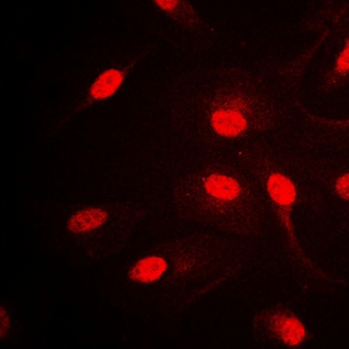
Immunofluorescent analysis of AP2 alpha/beta staining in HepG2 cells. Formalin-fixed cells were permeabilized with 0.1% Triton X-100 in TBS for 5-10 minutes and blocked with 3% BSA-PBS for 30 minutes at room temperature. Cells were probed with the primary antibody in 3% BSA-PBS and incubated overnight at 4 °C in a humidified chamber. Cells were washed with PBST and incubated with a DyLight 594-conjugated secondary antibody (red) in PBS at room temperature in the dark.

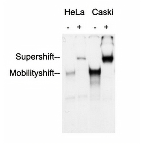
Anti-AP2 alpha/beta Antibody was used in an Electrophoretic Mobility Shift Assay (EMSA) to supershift the protein-DNA complex. Radiolabelled, double-stranded DNA oligonucleotides (10.000 cpm per lane) harbouring a binding site for AP2 alpha/beta were incubated with each 2 ug of nuclear extract (NE) from HeLa and Caski cells, respectively. Samples were incubated for 30 minutes at room temperature to allow the formation of protein-DNA complexes. Anti-AP2 alpha/beta Antibody were added to the samples (as indicated) and incubated for further 60 minutes at 4°C. Samples were separated in a 5.5% PAGE. The Gel was dried under vacuum and for autoradiography a X-ray film was exposed with an intensifying screen for 2 days at -80°C. Specific protein-DNA complexes were quantitatively supershifted with Anti-AP2 alpha/beta Antibody, verifying the binding of AP2 alpha/beta to the DNA oligonucleotide.

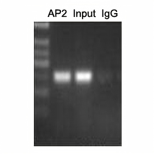
ChIP analysis of Cervical cancer cell lines lysate, incubated for 12 hours at 4°C. Cross-linking (X-ChIP) using formaldehyde for 10 minutes. Detection step: Semiquantitative PCR. Positive control: Tumor cell lines Hela. Negative control: Human primary keratinocytes.

Storage
Store at 4°C short term. For long term storage, store at -20°C, avoiding freeze/thaw cycles.
Note
For Research Use Only. Not for use in diagnostic procedures.
FAQs
What are the main types of research antibodies and how do they differ?
Research antibodies are mainly divided into monoclonal antibodies and polyclonal antibodies. Monoclonal antibodies typically offer higher specificity and better batch-to-batch consistency, while polyclonal antibodies often provide stronger affinity but may show more variation between batches. The choice depends on your specific experimental needs.
How can I tell if a research antibody is suitable for my experiment?
It is recommended to carefully review the product datasheet for validated applications, species reactivity, recommended dilutions, and published references. For new antibodies, performing a small-scale validation with positive control samples is usually helpful.
Can improper storage of research antibodies affect experimental results?
Yes. Antibodies are sensitive to temperature, repeated freeze-thaw cycles, and contamination. Improper storage may lead to reduced activity, increased background, or weaker signals. It is best to follow the storage instructions provided in the product datasheet.
Why doesn’t the recommended dilution in the datasheet work well in my experiment?
The recommended dilution is based on the supplier’s test conditions. Factors such as sample type, fixation method, and detection system in your lab can influence the optimal working concentration. Performing a dilution series optimization in your own system is often necessary.
What precautions should I take when using a newly purchased research antibody for the first time?
It is advisable to briefly centrifuge the antibody (especially concentrated or lyophilized ones), then perform a small-scale pilot experiment using the recommended conditions. Recording the batch number and usage date is also helpful for future tracking.
New Products
Get in touch with AREX
Name:*
Tel/Phone:*
Company:*
Email:*
Inquiry:
Captcha*
Submitting your email information means that you are willing to receive email information from AREX regarding technology, applications, products, and events. By clicking on the 'unsubscribe' button in the email or by contacting info@arexbio.com You can unsubscribe at any time by sending an email. Regarding your data usage information, please refer to our privacy policy.
© AREX 2024. All Rights Reserved. Sitemap
0.167988s