VEGFR3 Mouse Monoclonal Antibody(C3773)


Key features and details
- Target:
- Host:
- Reactivity:
- Clonality:
- Applications:
- Conjugation:
- Gene Symbol:
- Storage:
-
Brand:

CAT.NO. : AMA03385
US$ Please choose
US$ Please choose
 Product Details
Background
Vascular endothelial growth factor is a key regulator of blood vessel development in embryos and angiogenesis in adult tissues. Unlike VEGF, the related VEGFC stimulates the growth of lymphatic vessels through its specificl ymphatic endothelial receptor VEGFR-3. In 1998, Dumont showed that targeted inactivation of the VEGFR-3 gene in mice resulted in defective blood vessel development in early embryos. Vasculogenesis and angiogenesis occurred, but large vessels became abnormally organized with defective lumens, leading to fluid accumulation in the pericardial cavity and cardiovascular failure at embryonic day 9.5. Thus, VEGFR3 has an essential role in the development of theembryonic cardiovascular system before the emergence of the lymphatic vessels.
Application
|
Application |
Dilution Ratio |
|
WB |
1:500 - 1:1000 |
|
IHC |
1:100 - 1:200 |
|
FC |
1:20 - 1:100 |
Overview
|
Product Description |
Mouse monoclonal antibody to VEGFR3 |
|
Immunogen |
Recombinant fusion protein of human VEGFR3. The exact sequence is proprietary. |
|
Purification Method |
This antibody is purified through a protein G column. |
|
Clonality |
Monoclonal |
|
Form |
Mouse IgG2a. Liquid in PBS, pH 7.3, 30% glycerol, and 0.01% sodium azide. |
|
Gene Symbol |
FLT4 |
|
Alternative Names |
VEGFR3; Vascular endothelial growth factor receptor 3; VEGFR-3; Fms-like tyrosine kinase 4; FLT-4; Tyrosine-protein kinase receptor FLT4 |
|
Gene ID (Human) |
2324 |
|
Protein ID (Human) |
P35916 |
Data
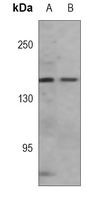
Western blot analysis of VEGFR3 expression in A549 (A), 293T (B) whole cell lysates. (Predicted band size: 152 kD; Observed band size: 153 kD)
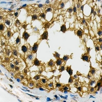
Immunohistochemical analysis of VEGFR3 staining in human testis formalin fixed paraffin embedded tissue section. The section was pre-treated using heat mediated antigen retrieval with sodium citrate buffer (pH 6.0). The section was then incubated with the antibody at room temperature and detected using an HRP conjugated compact polymer system. DAB was used as the chromogen. The section was then counterstained with haematoxylin and mounted with DPX.

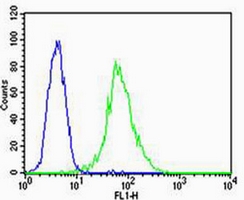
Flow cytometric analysis of HUVEC cells using Anti-VEGFR3 Antibody. The cells were fixed with 2% paraformaldehyde (10 min) and then permeabilized with 90% methanol for 10 min. The cells were incubated in 2% bovine serum albumin to block non-specific protein-protein interactions followed by the antibody at 37 °C for 60 min. The secondary antibody Goat Anti-Mouse IgG (H&L) - AF488 was incubated at 37 °C for 40 min. Isotype control antibody (blue line) was used under the same condition.
Storage
Store at +4℃ after thawing. Aliquot store at -20℃. Avoid repeated freeze / thaw cycles.
Note
For Research Use Only. Not for use in diagnostic procedures.
New Products