ERK1/2 kanin polyklonal antikropp
 MSDS
MSDS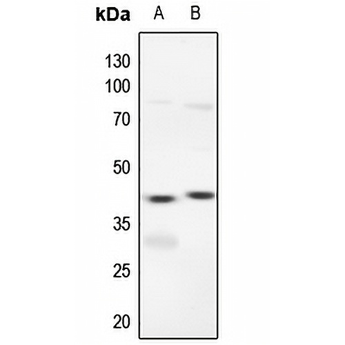
Nyckelfunktioner och detaljer
- Mål:
- Källa/värd:
- Reaktivitet:
- Klonalitet:
- Applikationer:
- Konjugering:
- Lagring:
-
Märke:

 Produktdetaljer
WB | 1:500 - 1:1000 |
IHC | 1:50 - 1:100 |
IF/ICC | 1:50 - 1:200 |
Beskrivning | Kanin polyklonal antikropp mot ERK1/2 |
Specificitet | Känner igen endogena nivåer av ERK1/2-protein. |
Antikroppstyp | Primär antikropp |
Imnunogen | KLH-konjugerad syntetisk peptid som omfattar en sekvens inom mittområdet av human ERK1/2. Den exakta sekvensen är proprietär. |
Rening | Antikroppen renades genom immunogenaffinitetskromatografi. |
Molekylvikt | Förutspått: 43; Observerad: 44 kD |
Form/buffert | Vätska i 0,42 % kaliumfosfat, 0,87 % natriumklorid, pH 7,3, 30 % glycerol och 0,01 % natriumazid. |
Alternativa namn | MAPK3; ERK1; PRKM3; Mitogen-activated protein kinase 3; MAP kinase 3; MAPK 3; ERT2; Extracellular signal-regulated kinase 1; ERK-1; Insulin-stimulated MAP2 kinase; MAP kinase isoform p44; p44-MAPK; Microtubule-associated protein 2 kinase; p44-ERK1; MAPK1; ERK2; PRKM1; PRKM2; Mitogen-activated protein kinase 1; MAP kinase 1; MAPK 1; ERT1; Extracellular signal-regulated kinase 2; ERK-2; MAP kinase isoform p42; p42-MAPK; Mitogen-activated protein kinase 2; MAP kinase 2; MAPK 2 |
Gensymbol | MAPK1; MAPK3 |
Entrez Gene | 5595; 5594(Människa); 50689; 116590(råtta) |
SwissProt | P27361; P28482 (Human); P21708; P63086(råtta) |
*Klonnummer, reaktivitet, källa/värd och klonalitet finns i avsnittet produktnamn och nyckelfunktioner ovan.
Western blot analysis of ERK1/2 expression in mouse lung (A), rat lung (B) whole cell lysates. (Predicted band size: 43; 41 kD; Observed band size: 44 kD)
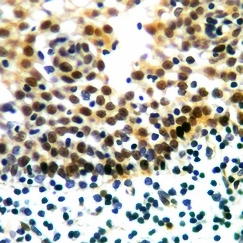
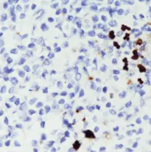
Immunohistochemical analysis of ERK1/2 staining in human tonsil cancer formalin fixed paraffin embedded tissue section. The section was pre-treated using heat mediated antigen retrieval with sodium citrate buffer (pH 6.0). The section was then incubated with the antibody at room temperature and detected using an HRP conjugated compact polymer system. DAB was used as the chromogen. The section was then counterstained with haematoxylin and mounted with DPX.
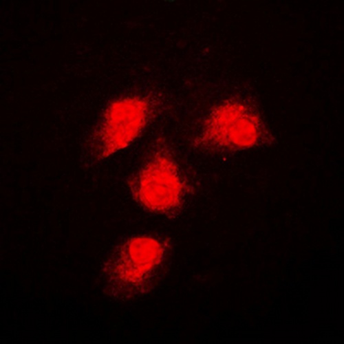
Immunofluorescent analysis of ERK1/2 staining in NIH3T3 cells. Formalin-fixed cells were permeabilized with 0.1% Triton X-100 in TBS for 5-10 minutes and blocked with 3% BSA-PBS for 30 minutes at room temperature. Cells were probed with the primary antibody in 3% BSA-PBS and incubated overnight at 4 °C in a humidified chamber. Cells were washed with PBST and incubated with a DyLight 594-conjugated secondary antibody (red) in PBS at room temperature in the dark.
Vanliga frågor
 Nya produkter