NRAS Rabbit Monoclonal Antibody(C4066)


Key features and details
- Reactivity:
- Application:
- Host:
- Clonality:
- Target:
-
Brand:

CAT.NO. : AMA03886
US$ Please choose
US$ Please choose
 Product Details
Background
NRAS (GTPase NRas) is a membrane protein that shuttles between the Golgi apparatus and the plasma membrane. This shuttling is regulated through palmitoylation and depalmitoylation by the ZDHHC9-GOLGA7 complex. This protein, which has intrinsic GTPase activity, is activated to a GTP-bound form by a GTPase activating protein and inactivated to a GDP-bound form by a guanine nucleotide-exchange factor. Defects in the gene encoding this protein are a cause of juvenile myelomonocytic leukemia (JMML) and Noonan syndrome 6.
Application
|
Application |
Dilution Ratio |
|
WB |
1:500-1:1000 |
|
IHC |
1:50-1:200 |
|
IF/ICC |
1:50-1:200 |
|
IP |
1:10-1:50 |
Overview
|
Product Description |
Recombinant rabbit monoclonal antibody to NRAS |
|
Immunogen |
KLH-conjugated synthetic peptide encompassing a sequence within human NRAS protein. The exact sequence is proprietary. |
|
Purification Method |
The antibody was purified by immunogen affinity chromatography. |
|
Clonality |
Monoclonal |
|
Form |
Liquid in PBS, pH 7.3, 50% glycerol, 0.05% BSA, and 0.05% Proclin300. |
|
Gene Symbol |
NRAS |
|
Alternative Names |
HRAS1; GTPase NRas; Transforming protein N-Ras |
|
Gene ID (Human) |
4893 |
|
Gene ID (Rat) |
24605 |
|
Protein ID (Human) |
P01111 |
|
Protein ID (Mouse) |
P08556 |
|
Protein ID (Rat) |
Q04970 |
Data
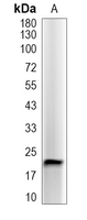
Western blot analysis of NRAS expression in Jurkat (A) whole cell lysates. (Predicted band size: 22 kD; Observed band size: 22 kD)
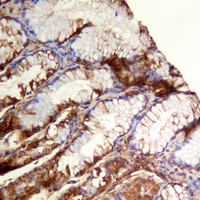
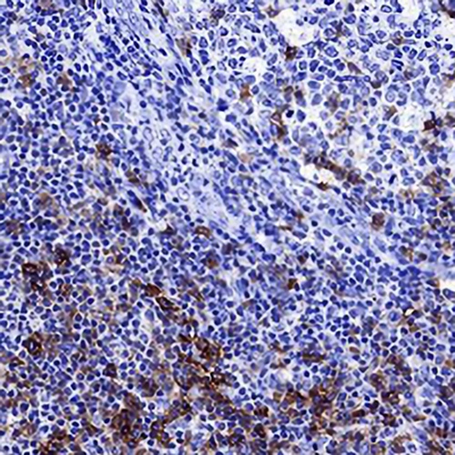
Immunohistochemical analysis of NRAS staining in human colon formalin fixed paraffin embedded tissue section. The section was pre-treated using heat mediated antigen retrieval with sodium citrate buffer (pH 6.0). The section was then incubated with the antibody at room temperature and detected using an HRP conjugated compact polymer system. DAB was used as the chromogen. The section was then counterstained with haematoxylin and mounted with DPX.
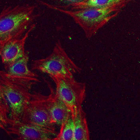
Immunofluorescent analysis of NRAS staining in Jurkat cells. Formalin-fixed cells were permeabilized with 0.1% Triton X-100 in TBS for 5-10 minutes and blocked with 3% BSA-PBS for 30 minutes at room temperature. Cells were probed with the primary antibody in 3% BSA-PBS and incubated overnight at 4 °C in a hidified chamber. Cells were washed with PBST and incubated with an AREX®Flour 488-conjugated secondary antibody (green) in PBS at room temperature in the dark. Phalloidin - AREX®Flour 594 was used to stain Actin filaments (red). DAPI was used to stain the cell nuclei (blue).
Storage
Store at 4°C short term. For long term storage, store at -20°C, avoiding freeze/thaw cycles.
Research Use Only
For Research Use Only. Not for use in diagnostic procedures.
New Products